

The pathologic characteristics of pingueculae on autofluorescence images
- PMID: 24311926
- PMCID: PMC3849304
- DOI: 10.3341/kjo.2013.27.6.416
The pathologic characteristics of pingueculae on autofluorescence images
Abstract
Purpose: To analyze the autofluorescence (AF) properties of pinguecula using cobalt-blue and yellow filters and to investigate the nature and pathogenesis of pingueculae using histochemical and immunohistochemical staining.
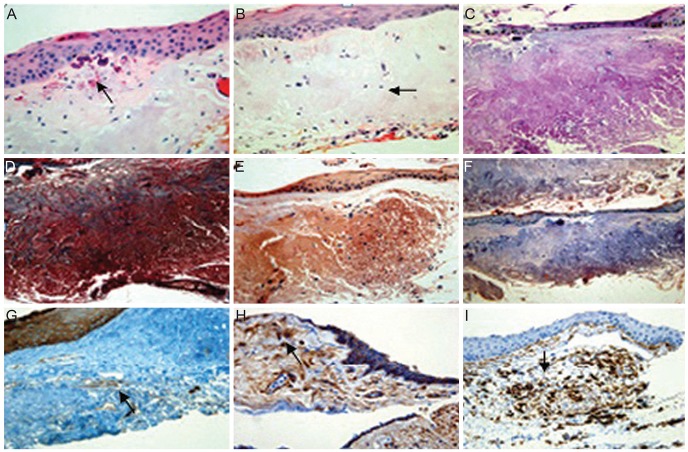
Methods: Fifty pingueculae in 40 patients were included in this study. AF of the pingueculae was observed and analyzed using a cobalt-blue filter with an additional yellow filter on a slit-lamp. Hematoxylin-eosin and immunohistochemical stainings were performed on surgical specimens of pingueculae that were prepared from each patient. Immunohistochemical staining included Congo red, Oil Red O, periodic acid-Schiff (PAS), Masson's trichrome, transglutaminase-2 (TG-2), mesenchymal stem cell markers CD29 (β-1-integrin), and CD34.
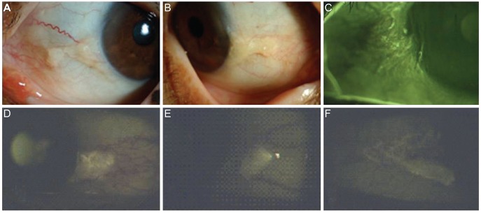
Results: AF images revealed hyper-AF in the pinguecula area. The AF lesions of pingueculae showed superficial punctuate erosions and avascular lesions. Deposition of eosinophilic and amorphous materials in the subepithelial layer of the pinguecula were observed on hematoxylin-eosin staining. Historeactivities to Congo red, PAS, Oil Red O, alcian blue, and Masson's trichrome were not detected, but immunoreactivities to CD29, CD34, and TG-2 were detected in the pingueculae with AF. However, CD29, CD34, and TG-2 were not detected in the pingueculae without AF.
Conclusions: The AF of pingueculae may be related to CD29, CD34, and TG-2. We suggest that pingueculae with AF have a different pathogenesis compared to pingueculae without AF.
Keywords: Optical imaging; Pinguecula; Transglutaminase 2.
Conflict of interest statement
No potential conflict of interest relevant to this article was reported.
Figures
Similar articles
-
Clinical and Autofluorescence Findings in Eyes with Pinguecula and Pterygium.J Ophthalmic Vis Res. 2023 Jul 28;18(3):260-266. doi: 10.18502/jovr.v18i3.13773. eCollection 2023 Jul-Sep. J Ophthalmic Vis Res. 2023. PMID: 37600917 Free PMC article.
-
Autofluorescence imaging of pingueculae.Br J Ophthalmol. 2009 Mar;93(3):396-9. doi: 10.1136/bjo.2008.144055. Epub 2008 Nov 19. Br J Ophthalmol. 2009. PMID: 19019934
-
Spectral domain anterior segment optical coherence tomography assessment of pterygium and pinguecula.Acta Ophthalmol. 2012 Aug;90(5):461-5. doi: 10.1111/j.1755-3768.2010.01994.x. Epub 2010 Oct 7. Acta Ophthalmol. 2012. PMID: 21040504
-
P53 expression in altered limbal basal cells of pingueculae, pterygia, and limbal tumors.Curr Eye Res. 1997 Dec;16(12):1179-92. doi: 10.1076/ceyr.16.12.1179.5036. Curr Eye Res. 1997. PMID: 9426949 Review.
-
The wonderful colors of the hematoxylin-eosin stain in diagnostic surgical pathology.Int J Surg Pathol. 2014 Feb;22(1):12-32. doi: 10.1177/1066896913517939. Epub 2014 Jan 9. Int J Surg Pathol. 2014. PMID: 24406626 Review.
Cited by
-
Clinical and Autofluorescence Findings in Eyes with Pinguecula and Pterygium.J Ophthalmic Vis Res. 2023 Jul 28;18(3):260-266. doi: 10.18502/jovr.v18i3.13773. eCollection 2023 Jul-Sep. J Ophthalmic Vis Res. 2023. PMID: 37600917 Free PMC article.
References
-
- Young JD, Finlay RD. Primary spheroidal degeneration of the cornea in Labrador and northern Newfoundland. Am J Ophthalmol. 1975;79:129–134. - PubMed
-
- Dushku N, Reid TW. P53 expression in altered limbal basal cells of pingueculae, pterygia, and limbal tumors. Curr Eye Res. 1997;16:1179–1192. - PubMed
-
- Dong N, Li W, Lin H, et al. Abnormal epithelial differentiation and tear film alteration in pinguecula. Invest Ophthalmol Vis Sci. 2009;50:2710–2715. - PubMed
-
- Spaide RF. Fundus autofluorescence and age-related macular degeneration. Ophthalmology. 2003;110:392–399. - PubMed
-
- Tatlpnar S, Ayata A, Unal M, Ersanl D. Fundus autofluorescence in choroidal rupture. Retin Cases Brief Rep. 2008;2:231–233. - PubMed
MeSH terms
Substances
LinkOut - more resources
Full Text Sources
Other Literature Sources
Miscellaneous
