

Case Reports
doi: 10.3341/kjo.2013.27.6.470.
Epub 2013 Nov 15.
Ophthalmic artery aneurysm: potential culprit of central retinal artery occlusion
Affiliations
- PMID: 24311936
- PMCID: PMC3849314
- DOI: 10.3341/kjo.2013.27.6.470
Item in Clipboard
Case Reports
Ophthalmic artery aneurysm: potential culprit of central retinal artery occlusion
Korean J Ophthalmol.
2013 Dec.
Abstract
Central retinal artery occlusion (CRAO) is one of the most devastating ophthalmic emergencies, causing acute painless visual loss in the affected eye. We describe the first case of acute non-arteritic CRAO associated with peripheral ophthalmic artery aneurysm and its clinical course after intra-arterial thrombolysis therapy. This case suggests that ophthalmic artery aneurysm can be the cause of CRAO and should be included in the differential diagnosis of CRAO.
Keywords: Ophthalmic artery aneurysm; Retinal artery occlusion.
Conflict of interest statement
No potential conflict of interest relevant to this article was reported.
Figures
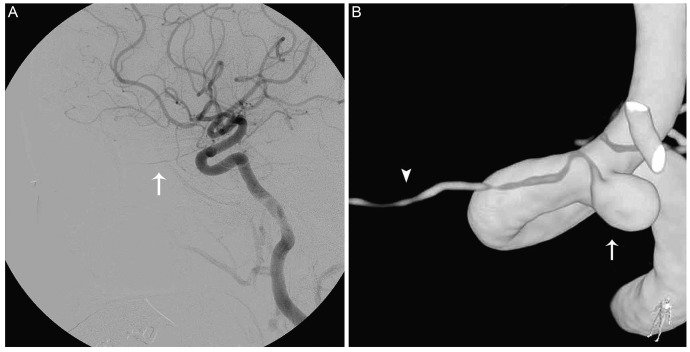
Internal carotid artery angiogram and three dimensional reconstructed image of the ophthalmic artery. (A) The internal carotid artery angiogram shows patent ophthalmic artery (arrow). (B) Three dimensional reconstructed view of the ophthalmic artery (arrow head) identifies its origin from the 3.7 × 4.5 × 5.2 mm sized aneurysm (arrow).
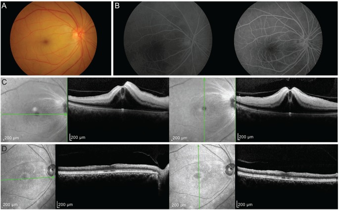
Fundus photography, fluorescein angiography and spectral-domain optical coherence tomography (SD-OCT) after intra-arterial thrombolysis. (A) One day after thrombolysis, the retina was still edematous with a typical "cherry red spot" appearance but showed improved vascularity. (B) Retinal arterial filling showed normal arterio-venous transit time of 11 seconds (left 20 seconds, right 31 seconds). (C) SD-OCT showed increased thickness and reflectivity of the inner retina typical of central retinal artery occlusion. (D) After 4 months of treatment, the inner retina thickness had markedly decreased.
Similar articles
-
Central Retinal Artery Occlusion Due to Intraorbital Ophthalmic Artery Aneurysm.J Neuroophthalmol. 2019 Mar;39(1):125-126. doi: 10.1097/WNO.0000000000000698. J Neuroophthalmol. 2019. PMID: 30052553 Review.
-
Analysis of the Effect of Superselective Ophthalmic Artery Thrombolysis for Central Retinal Artery Occlusion.Ophthalmic Res. 2024;67(1):387-392. doi: 10.1159/000539362. Epub 2024 Jun 19. Ophthalmic Res. 2024. PMID: 38897178
-
Successful intra-arterial thrombolysis for central retinal artery occlusion secondary to chronic internal carotid artery occlusion: a case report.BMC Ophthalmol. 2025 Jul 10;25(1):404. doi: 10.1186/s12886-025-04248-9. BMC Ophthalmol. 2025. PMID: 40640788 Free PMC article.
-
Intra-arterial thrombolysis for central retinal artery occlusion: two cases report.J Korean Med Sci. 2010 Jun;25(6):974-9. doi: 10.3346/jkms.2010.25.6.974. Epub 2010 May 24. J Korean Med Sci. 2010. PMID: 20514326 Free PMC article.
-
Sequential bilateral retinal artery occlusions with promising visual prognosis in a diabetic patient: a case report and literature review.BMC Ophthalmol. 2025 Jun 2;25(1):331. doi: 10.1186/s12886-025-04166-w. BMC Ophthalmol. 2025. PMID: 40457210 Free PMC article. Review.
Cited by
-
Sequential central retinal artery and posterior ciliary artery occlusion due to ophthalmic artery aneurysm.BMJ Case Rep. 2024 May 2;17(5):e257568. doi: 10.1136/bcr-2023-257568. BMJ Case Rep. 2024. PMID: 38697685 No abstract available.
-
Central retinal artery occlusion after endovascular coil embolization for internal carotid artery aneurysm.Int J Ophthalmol. 2019 Mar 18;12(3):520-522. doi: 10.18240/ijo.2019.03.26. eCollection 2019. Int J Ophthalmol. 2019. PMID: 30918825 Free PMC article. No abstract available.
-
Intra-Arterial Thrombolysis for Central Retinal Artery Occlusion.Clin Ophthalmol. 2019 Dec 13;13:2489-2509. doi: 10.2147/OPTH.S232560. eCollection 2019. Clin Ophthalmol. 2019. PMID: 31853171 Free PMC article. Review.
References
-
- Rumelt S, Dorenboim Y, Rehany U. Aggressive systematic treatment for central retinal artery occlusion. Am J Ophthalmol. 1999;128:733–738. - PubMed
-
- Qiao L, Wang H, Mao L, et al. Peripheral ophthalmic artery aneurysm. Neurosurg Rev. 2011;34:29–38. - PubMed
-
- Qureshi AI, Mohammad Y, Yahia AM, et al. Ischemic events associated with unruptured intracranial aneurysms: multicenter clinical study and review of the literature. Neurosurgery. 2000;46:282–289. - PubMed
-
- Haritoglou C, Muller-Schunk S, Weber C, et al. Central retinal artery occlusion in association with an aneurysm of the internal carotid artery. Am J Ophthalmol. 2001;132:270–271. - PubMed
Publication types
MeSH terms
Substances
LinkOut - more resources
Full Text Sources
Other Literature Sources
Medical
