. 2013 Dec;11(12):e1001725.
doi: 10.1371/journal.pbio.1001725. Epub 2013 Dec 3.

NK4 antagonizes Tbx1/10 to promote cardiac versus pharyngeal muscle fate in the ascidian second heart field

Affiliations

NK4 antagonizes Tbx1/10 to promote cardiac versus pharyngeal muscle fate in the ascidian second heart field

Wei Wang et al. PLoS Biol. 2013 Dec.

Abstract

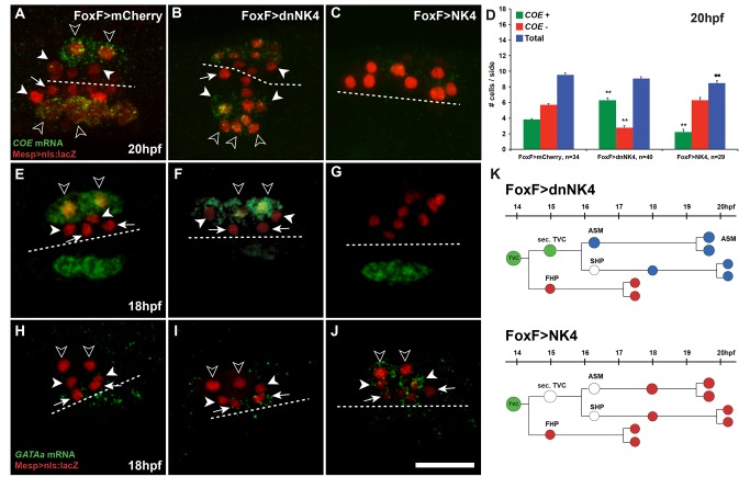
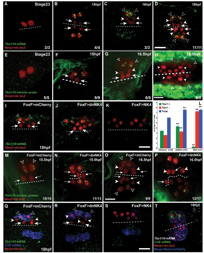
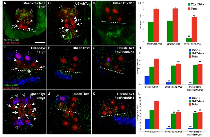
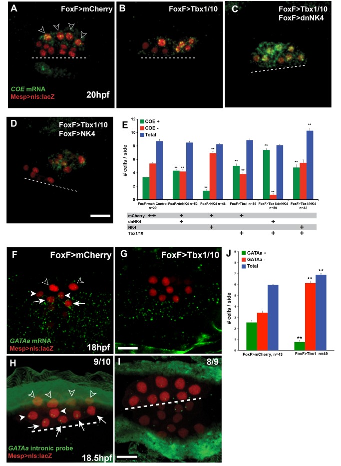
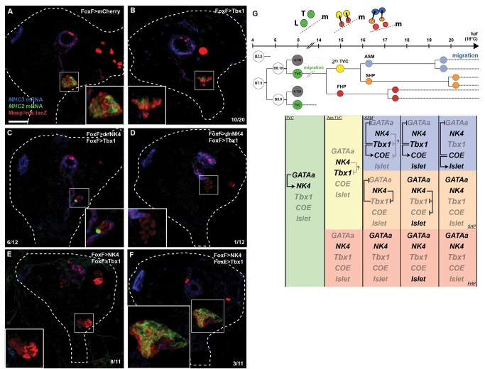
The heart and head muscles share common developmental origins and genetic underpinnings in vertebrates, including humans. Parts of the heart and cranio-facial musculature derive from common mesodermal progenitors that express NKX2-5, ISL1, and TBX1. This ontogenetic kinship is dramatically reflected in the DiGeorge/Cardio-Velo-Facial syndrome (DGS/CVFS), where mutations of TBX1 cause malformations in the pharyngeal apparatus and cardiac outflow tract. Cardiac progenitors of the first heart field (FHF) do not require TBX1 and segregate precociously from common progenitors of the second heart field (SHF) and pharyngeal muscles. However, the cellular and molecular mechanisms that govern heart versus pharyngeal muscle specification within this lineage remain elusive. Here, we harness the simplicity of the ascidian larva to show that, following asymmetric cell division of common progenitors, NK4/NKX2-5 promotes GATAa/GATA4/5/6 expression and cardiac specification in the second heart precursors by antagonizing Tbx1/10-mediated inhibition of GATAa and activation of Collier/Olf/EBF (COE), the determinant of atrial siphon muscle (ASM) specification. Our results uncover essential regulatory connections between the conserved cardio-pharyngeal factor Tbx1/10 and muscle determinant COE, as well as a mutual antagonism between NK4 and Tbx1/10 activities upstream of GATAa and COE. The latter cross-antagonism underlies a fundamental heart versus pharyngeal muscle fate choice that occurs in a conserved lineage of cardio-pharyngeal progenitors. We propose that this basic ontogenetic motif underlies cardiac and pharyngeal muscle development and evolution in chordates.


Conflict of interest statement

The authors have declared that no competing interests exist.

Figures

Figure 1
Figure 1. NK4 promotes heart formation at the expense of the ASMs and LoMs in Ciona intestinalis.
(A) Summary of early cardio-pharyngeal development: cell divisions, lineage, and migration of the TVCs. Green, TVCs and secondary TVCs (2ary TVC); red, FHPs and SHP; blue, ASMs and LoM precursors; OSMs, Oral Siphon Muscles; ATMs, anterior tail muscles; hpf, hours postfertilization. Green and blue arrows, TVC and ASM migrations, respectively. (B–D) 72 hpf juveniles electroporated with Mesp>nls:lacZ (red) to mark B7.5 lineage cells and FoxF>mCherry (B), FoxF>dnNK4 (C), or FoxF>NK4 (D). Double FISH-IHC with MHC2 (green) and MHC3 (blue) probes mark cardiomyocytes and ASM/LoMs, respectively. White dotted lines outline whole bodies. Insets show magnified hearts. Numbers indicate juveniles showing the phenotype and total. Scale bar, 25 µm.
Figure 2
Figure 2. NK4 represses COE and activates GATAa in TVC derivatives.
(A–C, E–G, H–J) Larvae electroporated with Mesp>nls:lacZ (red), FoxF>mCherry (A, E, H), FoxF>dnNK4 (B, F, I), and FoxF>NK4 (C, G, H). FISH of COE transcripts (green) at 20 hpf (A–C) and 18 hpf (E–G) and GATAa transcripts (green) at 18 hpf (H–J). White dotted lines indicate the midline. Target expression of –dnNK4 in TVC lineage induces ectopic COE expression in SHPs (white arrowheads), in addition to ASM precursors (open arrowheads), but not in the FHPs (B, F, arrows). Conversely, FoxF>NK4 represses COE expression (C, G). In wild-type embryos, endogenous GATAa expression is restricted to heart precursors (arrows, H). FoxF>dnNK4 does not affect GATAa expression in the FHP (arrows, I). In FoxF>NK4 expressing larvae, GATAa transcripts can be detected in all TVC derivatives, including the lateral-most (open arrowheads). Scale bar, 25 µm. (D) Numbers of βGal and COE expressing cells per electroporated half at 20 hpf, in mCherry controls, dnNK4, or NK4 expressing larvae. n, number of embryo halves scored. Error bar, standard error of the mean (SEM), Student's t test compared experimental condition to the control. **p<0.05. (K) Interpretation of TVC lineage fate re-programming by Fox>dnNK4 and FoxF>NK4. FoxF>dnNK4, SHPs are converted into ASMs; FoxF>NK4, ASMs are converted into heart precursors.
Figure 3
Figure 3. NK4 contributes to restricting Tbx1/10 expression to the ASM precursors.
(A–D, I–K and Q–S) Tbx1/10 mRNAs (green), (E–H, M–P) Tbx1/10 nascent transcripts (green) and (Q–S) COE mRNA (blue). Larvae electroporated with Mesp>nls:lacZ (red), FoxF>mCherry (I, M and O), FoxF>dnNK4 (J, N and P), and FoxF>NK4 (K). Ventral views, dotted lines indicate the midline; hpf, hours postfertilization; stage 23, 14 hfp at 16°C. Scale bar, 10 µm. Targeted expression of dnNK4 in the TVCs induces ectopic Tbx1/10 expression in the SHP, but not in the FHPs (J arrows). FoxF>NK4 represses Tbx1/10 expression in all TVC derivates (K). (L) Histograms showing the number of Tbx1+ and βGal+ cells per half. n, number of embryo halves scored. Student's t test compared experimental condition to the control. **p<0.05, Error bar, standard error of the mean (SEM). COE transcripts can only be detected in lateral Tbx1/10 positive cells in control larvae at 18 hpf (Q). –dnNK4 causes expansion of COE expression to the medial SHPs, which express Tbx1/10 (R). Both Tbx1/10 and COE expressions were inhibited by NK4 overexpression (S). (T) Larva (16 hpf) co-electroporated with Mesp>nls:lacZ (red, nuclei) and Mesp>LifeAct:mCherry (blue, cell cortex) and hybridized with COE (green) and Tbx1/10 (white) probes. Tbx1/10 transcripts were detected in both ASM and SHPs, but not in the FHPs.
Figure 4
Figure 4. RNAi-mediated loss of Tbx1/10 function inhibits COE and causes ectopic GATAa expression.
Open arrowheads, ASM precursors; white arrowheads, SHPs; arrows, FHPs. Dotted lines indicate the midline. (A–C) Larva (16 hpf) electroportated with Mesp>nls:lacZ (loading control) (A), U6>shTyr (shRNA targeting the pigment cell-specific Tyrosinase, used as negative control, B), and U6>shTbx1/10 (C). TVCs are labeled with Mesp>nls:lacZ (red) by immunostaining. U6>shTbx1/10 knocked down the endogenous Tbx1/10 transcripts and induced a delay in secondary TVC division. Double FISH detection of COE (blue) and GATAa (green) expression in larva of 19 hpf (E–G) and 20 hpf (I–K). ShRNA-mediated knock-down of Tbx1/10 caused ectopic activation of GATAa in the lateral-most TVCs, while COE expression was markedly down-regulated (F, J). Note the persistent expression of COE in an ASM that did not receive the plasmids (open arrowhead in J). The co-electroporation of FoxF>dnNK4 with U6>shTbx1/10 shows no ectopic COE expression, but a remarkable down-regulation in the lateral-most TVCs. Ectopic GATAa expression is detected in the lateral-most TVCs as well as in larvae electroporated with U6>shTbx1/10 only (G, K). Histograms showing the number of Tbx1/10+, COE+, GATAa+, and βGal+ cells per half (D, H, and L). n, number of embryo halves scored. Student's t test compared experimental condition to the control. **p<0.05; error bar, standard error of the mean (SEM).
Figure 5
Figure 5. Tbx1/10 activity promotes COE and inhibits GATAa expression in the secondary TVC derivatives.
(A–D, F–I) Larva electroporated with Mesp>nls:lacZ (red) and indicated combinations of FoxF>mCherry, FoxF>dnNK4, FoxF>NK4, and FoxF>Tbx1/10. FISH detection of COE mRNAs (green) at 20 hpf (A–D) and GATAa mRNAs (green) at 18 hpf (F, G) and GATAa nascent transcripts at 18.5 hpf (H, I). White dotted lines indicate the midline. ASM precursors, open arrowheads; SHPs, white arrowheads,;FHPs, arrows. Scale bar, 10 µm. In control larvae (FoxF>mCherry, A), COE expression is restricted to ASM precursors; with FoxF>Tbx1/10 (B), weak COE expression expanded to a subset of heart precursors. Combined FoxF>Tbx1/10 and FoxF>dnNK4 strongly activated COE expression in all TVC derivatives (C). FoxF>Tbx1/10 and FoxF>NK4 caused sporadic ectopic activation of COE (D). (E) Histograms showing the numbers of COE+ and βGal+ cells per electroporated half. n, number of embryo halves scored. Student's t test compared experimental conditions to the control. **p<0.05; error bar, standard error of the mean (SEM). In the control, endogenous GATAa expression is restricted to heart precursors (F, H). FoxF>Tbx1/10 inhibits GATAa expression in all heart precursors (G, I). Histogram (J), numbers of βGal and GATAa expressing cells per electroporated half at 18 hpf. Error bar, standard error of the mean (SEM); Student's t test compared experimental condition to the control. **p<0.05.
Figure 6
Figure 6. NK4 binds to the minimal ASM enhancer of COE.
(A) Combined snapshots of the ANISEED and VISTA browsers showing the KH2008 transcript models for COE and conservation between Ciona intestinalis and Ciona savignyi. Pink peaks indicate conserved noncoding sequences (>65% identity per 80 bp). (B) Alignment of the conserved noncoding region corresponding to the minimal ASM enhancer (Figure S8). Above sequence, C. intestinalis; bottom sequence, C. savignyi. Putative NK4 and Tbx1/10 sites are highlighted in green and blue, respectively. Coordinates are expressed relative to the translation start site (ATG, where A is +1). (C) ChIP-qPCR data expressed as average fold enrichment relative to the mock control (ChIP using nonspecific IgG on the chromatin extracted from larvae electroporated with FoxF>NK4:2xFLAG), for ChIP using an anti-FLAG tag antibody on chromatin samples obtained from larvae electroporated with GFP:2xFLAG, NK4:2xFLAG, or dnNK4:2xFLAG. Error bars, standard error of the mean (SEM), calculated over biological triplicates and qPCR performed with two primer pairs for COE and two pairs for the internal loading control Brachyury.
Figure 7
Figure 7. Combined Tbx1/10 mis-expression and NK4 inhibition can convert all TVC derivatives into differentiated ASMs and LoMs in 72 hpf juveniles.
(A–F) Juveniles electroporated with Mesp>nls:lacZ(red) and indicated constructs were hybridized with MHC3 (blue) and MHC2 (green) probes. (A) FoxF>mCherry control. (B) FoxF>Tbx1/10 reduced heart volume and the number of MHC2+ cells in 10/20 animals; (C–D) combined FoxF>dnNK4 and FoxF>Tbx1/10 converted most (C) or all (D) TVC derivatives into differentiated ASMs and LoMs; (E–F) combined FoxF>NK4 and FoxF>Tbx1/10 showed least dramatic effects than each one alone: ASM/LoM formation defects are observed, 8/11 juveniles have reduced hearts and MHC2 expression, and 3/11 juveniles have enlarged heart and increased MHC2 expression. White dotted lines outline whole bodies. Insets show magnified hearts. Scale bar, 25 µm. (G) Summary model of the NK4 and Tbx1/10 mutual antagonism regulating cardiac versus ASM fate specification within a conserved clonal topology for progressive fate choices. The cells and approximate time windows are showed (i.e., align with the timeline above). Greyed labels indicate inactive genes and regulatory interactions. The question marks point to the unknown mechanism(s) that prevent(s) NK4-mediated inhibition of Tbx1/10 and COE expression in the secondary TVCs and ASM founder cells.

Similar articles

Cited by

References

    1. Jerome LA, Papaioannou VE (2001) DiGeorge syndrome phenotype in mice mutant for the T-box gene, Tbx1. Nat Genet 27: 286–291. - PubMed
    1. Merscher S, Funke B, Epstein JA, Heyer J, Puech A, et al. (2001) TBX1 is responsible for cardiovascular defects in velo-cardio-facial/DiGeorge syndrome. Cell 104: 619–629. - PubMed
    1. Kelly RG, Jerome-Majewska LA, Papaioannou VE (2004) The del22q11.2 candidate gene Tbx1 regulates branchiomeric myogenesis. Hum Mol Genet 13: 2829–2840. - PubMed
    1. Lindsay EA, Vitelli F, Su H, Morishima M, Huynh T, et al. (2001) Tbx1 haploinsufficieny in the DiGeorge syndrome region causes aortic arch defects in mice. Nature 410: 97–101. - PubMed
    1. Nathan E, Monovich A, Tirosh-Finkel L, Harrelson Z, Rousso T, et al. (2008) The contribution of Islet1-expressing splanchnic mesoderm cells to distinct branchiomeric muscles reveals significant heterogeneity in head muscle development. Development 135: 647–657. - PMC - PubMed

Publication types

MeSH terms

LinkOut - more resources