Review
. 2013 Nov 19:7:182.
doi: 10.3389/fncir.2013.00182.

Vestibular blueprint in early vertebrates

Affiliations
Review

Vestibular blueprint in early vertebrates

Hans Straka et al. Front Neural Circuits. .

Abstract

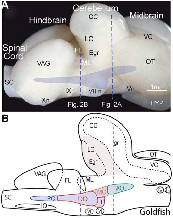
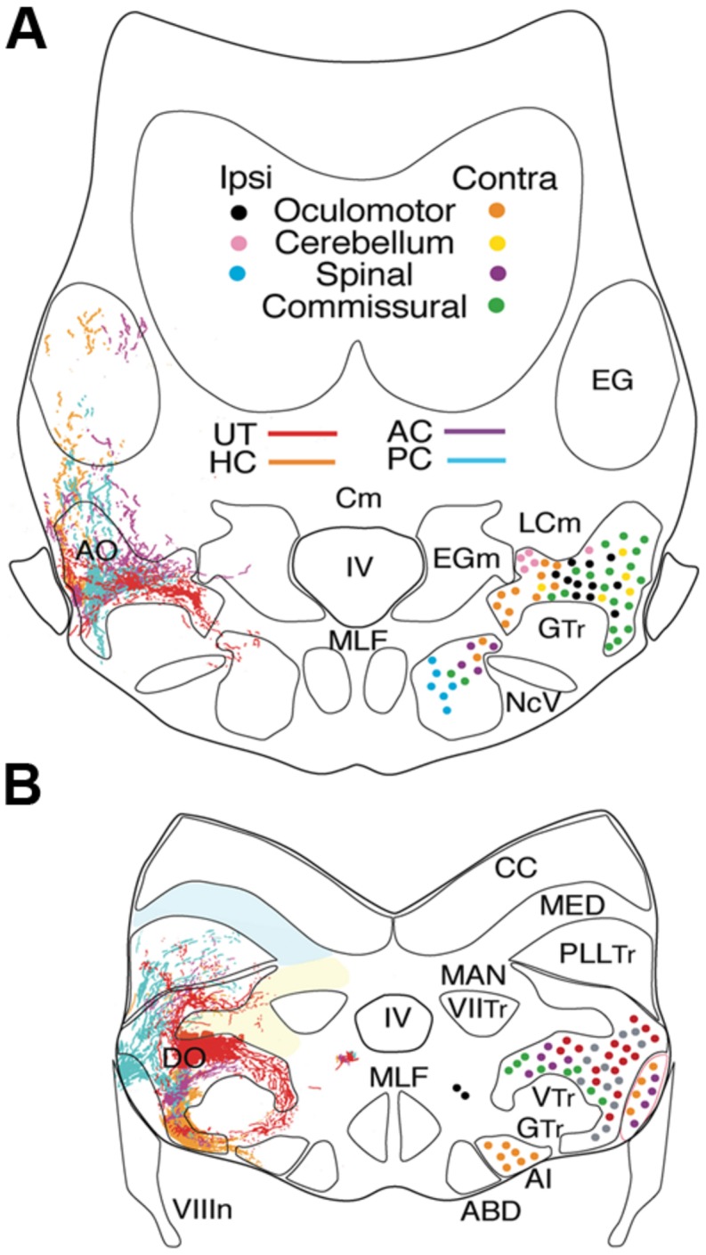
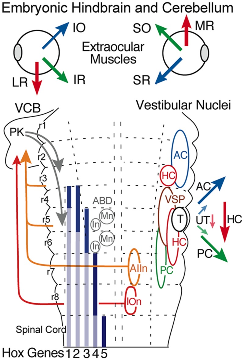
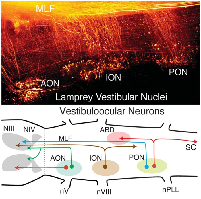
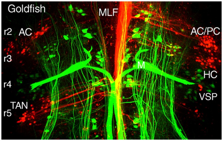
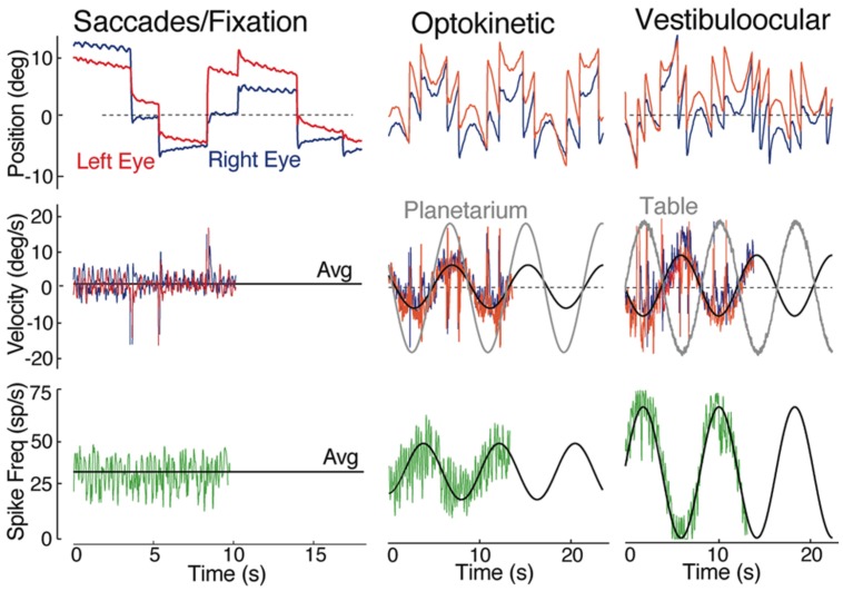
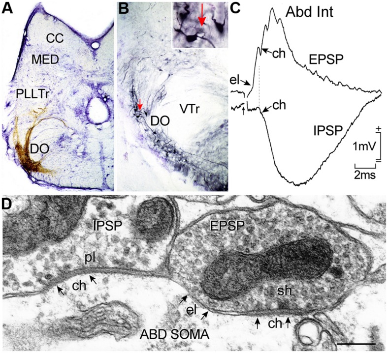
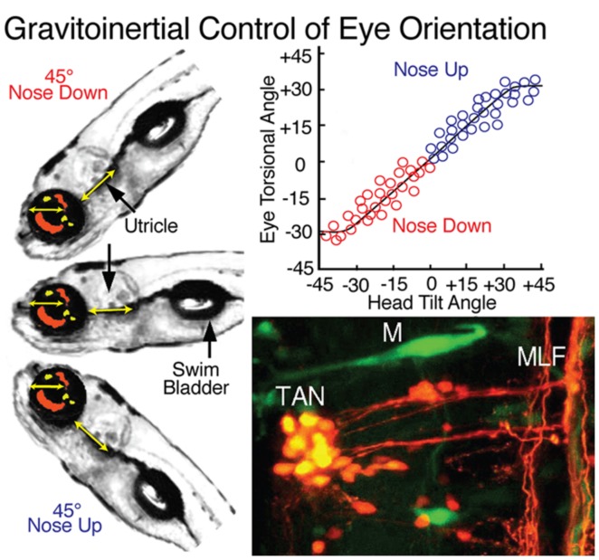
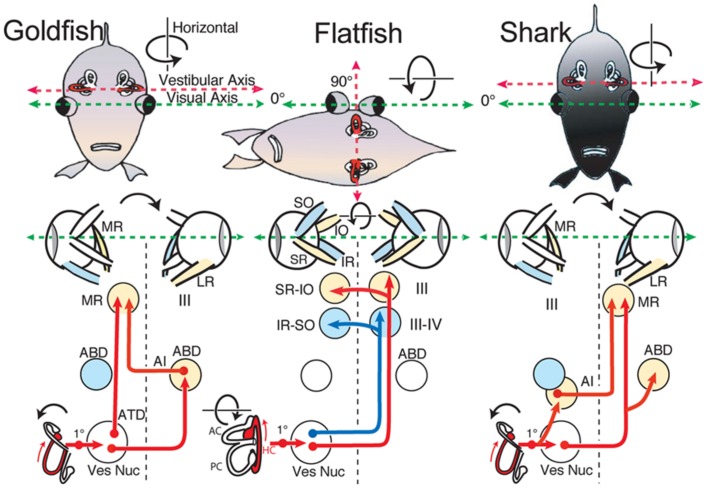
Central vestibular neurons form identifiable subgroups within the boundaries of classically outlined octavolateral nuclei in primitive vertebrates that are distinct from those processing lateral line, electrosensory, and auditory signals. Each vestibular subgroup exhibits a particular morpho-physiological property that receives origin-specific sensory inputs from semicircular canal and otolith organs. Behaviorally characterized phenotypes send discrete axonal projections to extraocular, spinal, and cerebellar targets including other ipsi- and contralateral vestibular nuclei. The anatomical locations of vestibuloocular and vestibulospinal neurons correlate with genetically defined hindbrain compartments that are well conserved throughout vertebrate evolution though some variability exists in fossil and extant vertebrate species. The different vestibular subgroups exhibit a robust sensorimotor signal processing complemented with a high degree of vestibular and visual adaptive plasticity.

Keywords: extraocular motoneurons; eye movements; goldfish; hindbrain segment; otolith; semicircular canal; vestibuloocular; vestibulospinal.


Figures

FIGURE 1
FIGURE 1
Location of the octavolateral vestibular subdivisions within the goldfish hindbrain. A side view of the intact brain (A) and sagittal schematic diagram (B) are drawn at the same scale. Vertical dashed lines indicate rostro-caudal levels of the coronal sections shown in Figure 2. AO, DO, MO, PO, anterior, descending medial, posterior octavolateral nuclei; CC, corpus cerebelli; Egr, external granule cell layer; FL, facial lobe; gr, corpus cerebelli granule cell layer; HYP, hypothalamus; III, IV, and VI oculomotor, trochlear, and abducens nuclei; VIIIn, IXn Xn, and Vn, vestibular, glossopharyngeal, vagal, and trigeminal nerves; IO, inferior olive; LC, lobus caudalis; ML, molecular layer of crus cerebelli; VC, valvula cerebelli; OT, optic tectum; SC, spinal cord; T, tangential nucleus; VAG, vagal lobe.
FIGURE 2
FIGURE 2
Afferent and efferent vestibular pathways in adult goldfish. Schematic cross sections at the level of r3 (A) and r5 (B) showing first order canal and utricular afferent terminations. Efferent projections to ipsi- and contralateral extraocular, spinal, cerebellar, and vestibular targets are color-coded. Reconstruction based on own data (Straka and Baker, unpublished) and published material (McCormick and Braford, 1994). AI, abducens internuclear neurons; ABD, abducens motoneurons; AO, DO, anterior, descending octavolateral nucleus; CC, cerebellar crest; EG, eminentia granularis; LC, caudal lobe; MAN, medial auditory nucleus; MED, lateral line medial nucleus; MLF, medial longitudinal fasciclus; T, tangential nucleus; GTr, secondary gustatory tract; PllTr, posterior lateral line tract; VTr, descending tract of the Vth cranial nerve; VIITr, central tract of the seventh cranial nerve.
FIGURE 3
FIGURE 3
Hindbrain segmental organization of vestibular nuclei with schematic diagram of canal and otolith-specific vestibuloocular behaviors and hox gene expression. Corresponding color-coded arrows and excitatory vestibuloocular subgroups in r1-3 and r5-7 indicate the close spatial alignment between canal planes and vertical, oblique, and horizontal extraocular muscle pulling directions. Afferents from the inferior olive (r8) and vestibular subgroups (r3, r5, r7) to the vestibulocerebellum (VCB) are necessary for vestibuloocular reflex plasticity. Adapted from Straka et al. (2001). ABD, abducens nucleus; AC, PC, HC, anterior vertical, posterior vertical, horizontal canal; In, internuclear neuron; IOn, inferior olivary neurons; IO, SO, inferior, superior oblique; IR, LR, MR, SR, inferior, lateral, medial, superior rectus; Mn, motoneuron; PK, Purkinje cell; T, tangential nucleus; UT, utricle; VSP, vestibulospinal.
FIGURE 4
FIGURE 4
Organization of lamprey octavolateral vestibular nuclei. Confocal image stack (top) showing retrogradely labeled vestibular neurons in three midbrain-projecting nuclei. Schematic diagram (bottom) of crossed and uncrossed vestibuloocular neurons in the anterior, inferior, posterior octavolateral nuclei (AON, ION, PON) projecting to motoneurons in the oculomotor (III), trochlear (IV), and abducens (ABD) nucleus. PON neurons (red lines) project to ABD Mns and the spinal cord (SC). Organizational scheme based on data reported by Gilland and Baker (1995) and Pombal et al. (1996). nV, trigeminal nerve; nPLL, posterior lateral line nerve; nVIII, vestibular nerve; MLF, medial longitudinal fasciculus.
FIGURE 5
FIGURE 5
Segmental organizations of vestibuloocular, vestibulospinal, and reticulospinal neurons in a young adult goldfish. A confocal image stack illustrates arrangements of canal-specific oculomotor/trochlear (red) in r2 (AC, PC), r3 (HC), r5 (TAN), and spinal cord-projecting neurons (green) in r4-5 adjacent to the large Mauthner cells in r4. Adapted from Suwa et al. (1996).
FIGURE 6
FIGURE 6
Spontaneous, visual and vestibular-evoked horizontal eye movements in adult goldfish. Eye position (top row) and eye velocity (middle row) of the right (blue trace) and left eye (red trace) with the correlated discharge of a vestibular neuron (bottom row) during spontaneous saccades with periods of fixation, horizontal sinusoidal optokinetic stimulation (planetarium), and vertical axis sinusoidal head rotation (vestibuloocular); the black line indicates the average (Avg) eye velocity (middle row); and neuronal firing rate. Data were adapted from Green et al. (1997).
FIGURE 7
FIGURE 7
Horizontal canal termination, second order vestibular neuron location and morpho-physiological correlates of excitatory and inhibitory vestibular inputs to abducens motoneurons and AI neurons. Nissl-stained transverse section through a goldfish hindbrain illustrating biocytin-labeled horizontal canal afferent terminations (A) and vestibular neurons in the DO (B) that were retrogradely labeled from the contralateral ABD nucleus; a labeled neuron (red arrow) in (B) is shown at higher magnification in the inset. Crossed EPSP and uncrossed IPSP in an abducens internuclear neuron following electrical stimulation of the horizontal semicircular canal nerve on both sides, respectively (C); responses are mediated by electrical (el); and/or chemical synapses (ch) between inhibitory (pl) and excitatory vestibular (sh) axonal terminations on abducens neurons as illustrated in the electronmicroscopic image (D). CC, cerebellar crest; DO, descending octavolateral nucleus; MED, lateral line medial nucleus; pl, pleiotropic; PllTr, posterior lateral line tract; sh, spheroidal; VTr, descending tract of the Vth cranial nerve; VTr, descending tract of the Vth cranial nerve. Data in (C,D) adapted from Graf et al. (1997) and Green et al. (1997).
FIGURE 8
FIGURE 8
Gravitoinertial control of eye orientation in larval zebrafish. Compensatory static eye positions attained during 45 nose up and -45 nose down pitch are illustrated by the fish insets on the left. Precise counter-rotation of the eyes (top right) derived solely from sensorimotor transformation of utricular signals in tangential (TAN) neurons (bottom right). M, Mauthner cell; MLF, medial longitudinal fascicle. Figure summarizes results from Moorman et al. (1999); Suwa et al. (1999) and Bianco et al. (2012).
FIGURE 9
FIGURE 9
Schematic diagram of horizontal vestibuloocular reflex circuitry in teleosts and elasmobranchs. “Goldfish” shows the principal three-neuronal vestibuloocular reflex linking the horizontal semicircular canal with contralateral abducens (ABD) and ipsilateral MR motoneurons. ”Flatfish” shows that after 90 displacement of the vestibular relative to visual axis (metamorphosis) compensatory eye movements are produced by redirecting horizontal canal signals to vertical and oblique (SR-IO and IR-SO) motoneurons. In “Shark,” horizontal canal/second order neurons project to contralateral ABD and MR motoneurons including ipsilateral AI neurons. 1, first order vestibular neuron; ATD, Ascending Tract of Deiter’s. Schematic diagrams based on results from Graf et al. (; goldfish), Graf and Baker (,b; flatfish), and Graf et al. (; shark).

References

    1. Baker R., Precht W., Llinás R. (1972). Cerebellar modulatory action on the vestibulo-trochlear pathway in the cat. Exp. Brain Res. 15 364–385 10.1007/BF00234124 - DOI - PubMed
    1. Baker R., Gilland E. (1996). “The evolution of hindbrain visual and vestibular innovations responsible for oculomotor function,” in The Acquisition of Motor Behavior in Vertebrates eds Bloedel J. R., Ebner T. J., Wise S. P. (Cambridge, MA: MIT Press; ) 29–55
    1. Baker R. (1998). From genes to behavior in the vestibular system. Otolaryngol. Head Neck Surg. 119 263–275 10.1016/S0194-5998(98)70061-0 - DOI - PubMed
    1. Beck J. C., Gilland E., Tank D. W., Baker R. (2004). Quantifying the ontogeny of optokinetic and vestibuloocular behaviors in zebrafish, medaka, and goldfish. J. Neurophysiol. 92 3546–3561 10.1152/jn.00311.2004 - DOI - PubMed
    1. Beck J. C., Rothnie P., Straka H., Wearne S. L., Baker R. (2006). Precerebellar hindbrain neurons encoding eye velocity during vestibular and optokinetic behavior in the goldfish. J. Neurophysiol. 96 1370–1382 10.1152/jn.00335.2006 - DOI - PubMed

Publication types

LinkOut - more resources