Review
. 2013 Nov 20:4:385.
doi: 10.3389/fimmu.2013.00385.

Human immunodeficiency virus and heparan sulfate: from attachment to entry inhibition

Affiliations
Review

Human immunodeficiency virus and heparan sulfate: from attachment to entry inhibition

Bridgette J Connell et al. Front Immunol. .

Abstract

By targeting cells that provide protection against infection, HIV-1 causes acquired immunodeficiency syndrome. Infection starts when gp120, the viral envelope glycoprotein, binds to CD4 and to a chemokine receptor usually CCR5 or CXCR4. As many microorganisms, HIV-1 also interacts with heparan sulfate (HS), a complex group of cell surface associated anionic polysaccharides. It has been thought that this binding, occurring at a step prior to CD4 recognition, increases infectivity by pre-concentrating the virion particles at the cell surface. Early work, dating from before the identification of CCR5 and CXCR4, showed that a variety of HS mimetics bind to the gp120 V3 loop through electrostatic interactions, compete with cell surface associated HS to bind the virus and consequently, neutralize the infectivity of a number of T-cell line-adapted HIV-1 strains. However, progress made to better understand HIV-1 attachment and entry, coupled with the recent identification of additional gp120 regions mediating HS recognition, have considerably modified this view. Firstly, the V3 loop from CXCR4-using viruses is much more positively charged compared to those using CCR5. HS inhibition of cell attachment is thus restricted to CXCR4-using viruses (such as T-cell line-adapted HIV-1). Secondly, studies aiming at characterizing the gp120/HS complex revealed that HS binding was far more complex than previously thought: in addition to the V3 loop of CXCR4 tropic gp120, HS interacts with several other cryptic areas of the protein, which can be induced upon CD4 binding, and are conserved amongst CCR5 and CXCR4 viruses. In view of these data, this review will detail the present knowledge on HS binding to HIV-1, with regards to attachment and entry processes. It will discuss the perspective of targeting the gp120 co-receptor binding site with HS mimetic compounds, a strategy that recently gave rise to entry inhibitors that work in the low nanomolar range, independently of co-receptor usage.

Keywords: CCR5/CXCR4; HIV-1; V3 loop; attachment and entry inhibition; co-receptor binding site; glycosaminoglycan; gp120; heparan sulfate.


Figures

Figure 1
Figure 1
Heparan sulfate structure. HS, whose biosynthesis is initiated by the attachment of xylose (star) to specific serine residues in HSPG core proteins, followed by the formation of a linking tetrasaccharide (xylose-galactose-galactose-glucuronic acid), is initially polymerized by an enzyme complex composed of Ext1 and Ext2 as a GlcA-GlcNAc repeat (black). In restricted regions, called S-domains (shown in red), the chain is extensively modified by a series of enzymatic reactions that remove the acetyl group from GlcNAc residues and substitute the resulting free amino groups with sulfates, epimerizes the adjacent GlcA into L-iduronic acid (IdoA) and adds sulfates on various positions: the C2 of the IdoA (and less frequently that of the GlcA), the C6 of the GlcNS (and less frequently that of the GlcNac), and finally at the C3 of GlcNS or GlcN units. Altogether, these modifications can generate (the theoretical number of) 48 different disaccharides, whose combination within the S-domain gives rise to a large diversity of structures and make up binding sites for protein ligands, as depicted for example with a model of a CXCL12-HS complex [from Ref. (21)].
Figure 2
Figure 2
In cis and in trans capture of HIV-1 by heparan sulfate. HS can play multiple roles during viral infection. (A) On top of cells that express large amount of HS, but low CD4, such as macrophages, HS can capture viral particles and facilitate in cis subsequent interaction with specific entry receptors. (B) HS from non-permissive cells such as endothelia or epithelia can sequester HIV-1 and then mediate in trans infection by presenting the virus to permissive cells. (C) HS can contribute to both attachment and transcytosis of HIV-1 through epithelia.
Figure 3
Figure 3
HIV-1 entry mechanism. (A) Schematic representation of the multi-step process of HIV-1 entry; from attachment to CD4 (left) to fusion between the viral and the cell membrane (right). The gp120 trimer, upon binding to CD4 (in green), experiences extensive structural changes that open up the variable loops V1/V2 and V3 (orange and yellow), and concomitantly expose and/or fold the so called CD4 induced bridging sheet that will be recognized by the co-receptor (CCR5 and/or CXCR4). This second interaction triggers the insertion of the gp41 fusion peptide into the cell membrane and promotes viral entry (Reprinted from Ref. (102), with permission from Elsevier). (B) Three-dimensional structure of gp120 in the CD4-bound conformation (from pdb:2b4c), showing the inner and outer domains, the V1/V2 loop stem, and the four β strands (CD4 induced bridging sheet in blue) that together with the V3 loop (in green) contribute to co-receptor selectivity and interaction.
Figure 4
Figure 4
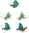
The V3 loop and the co-receptor binding domain of gp120 features HS binding sites. (A) The gp120 CD4-induced domain displays a HS binding structure. The Connolly surface of the HXBc2 gp120 core was color-coded according to the electrostatic potential from negative values (blue) to positive values (red). The basic residues of the CD4-induced (CD4i) epitope, which form a HS binding site, are indicated. (B) Representation of the lowest energy model of a gp120/HS derived octasaccharide complex. The protein [orientation as in (A)] is represented by a ribbon, and the octasaccharide by sticks. (C) Lowest energy model of the gp120 (on which the Connolly surface was calculated) in complex with a HS derived octasaccharide. V1/V2 and V3 indicate the stem of the V1/V2 and V3 loops. The location of the CD4i epitope is also indicated. (D) Structure of gp120, on which the V3 loop (in blue) was modeled. The HS binding residues of the V3 loop and the CD4-induced epitope are aligned on the surface of the protein and form an extended binding site on which has been docked a HS derived oligosaccharide of appropriate length. (E) Connolly surface of gp120, including the V3 loop, in complex with a tetradecasaccharide shown with the same orientation as in (C).
Figure 5
Figure 5
Principe of inhibition of HIV-1 attachment and entry by “CD4-HS.” (A) A CD4 mimetic peptide covalently linked to a HS dodecassaccharide (CD4-HS) bind to gp120 through its CD4 moiety and exposes the CD4i epitope, which then becomes available for interaction with the oligosaccharide. Such a bivalent molecule simultaneously binds to the CD4, the HS and the co-receptor binding sites of gp120 and blocks the interaction of the virus with all its principal cell surface ligands, inhibiting both attachment and entry (The gp120 was schematically represented as in Figure 3). (B) The structure of the mCD4-HS12 is also shown [modified from Ref. (99)].

References

    1. Barre-Sinoussi F, Chermann JC, Rey F, Nugeyre MT, Chamaret S, Gruest J, et al. Isolation of a T-lymphotropic retrovirus from a patient at risk for acquired immune deficiency syndrome (AIDS). Science (1983) 220:868–7110.1126/science.6189183 - DOI - PubMed
    1. Weiss RA. HIV receptors and cellular tropism. IUBMB Life (2002) 53:201–510.1080/15216540212652 - DOI - PubMed
    1. Shaw GM, Hunter E. HIV transmission. Cold Spring Harb Perspect Med (2012) 2(11):a006965.10.1101/cshperspect.a006965 - DOI - PMC - PubMed
    1. Berger EA, Murphy PM, Farber JM. Chemokine receptors as HIV-1 coreceptors: roles in viral entry, tropism, and disease. Annu Rev Immunol (1999) 17:657–70010.1146/annurev.immunol.17.1.657 - DOI - PubMed
    1. Ugolini S, Mondor I, Sattentau QJ. HIV-1 attachment: another look. Trends Microbiol (1999) 7:144–910.1016/S0966-842X(99)01474-2 - DOI - PubMed