. 2013 Nov 28;8(11):e77756.
doi: 10.1371/journal.pone.0077756. eCollection 2013.

Detection of HIV-1 neutralizing antibodies in a human CD4⁺/CXCR4⁺/CCR5⁺ T-lymphoblastoid cell assay system

Affiliations

Detection of HIV-1 neutralizing antibodies in a human CD4⁺/CXCR4⁺/CCR5⁺ T-lymphoblastoid cell assay system

Robert J McLinden et al. PLoS One. .

Abstract

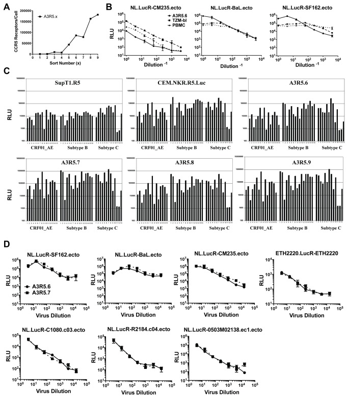
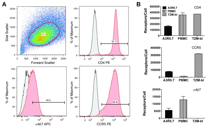
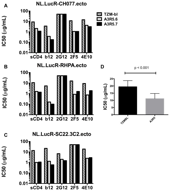
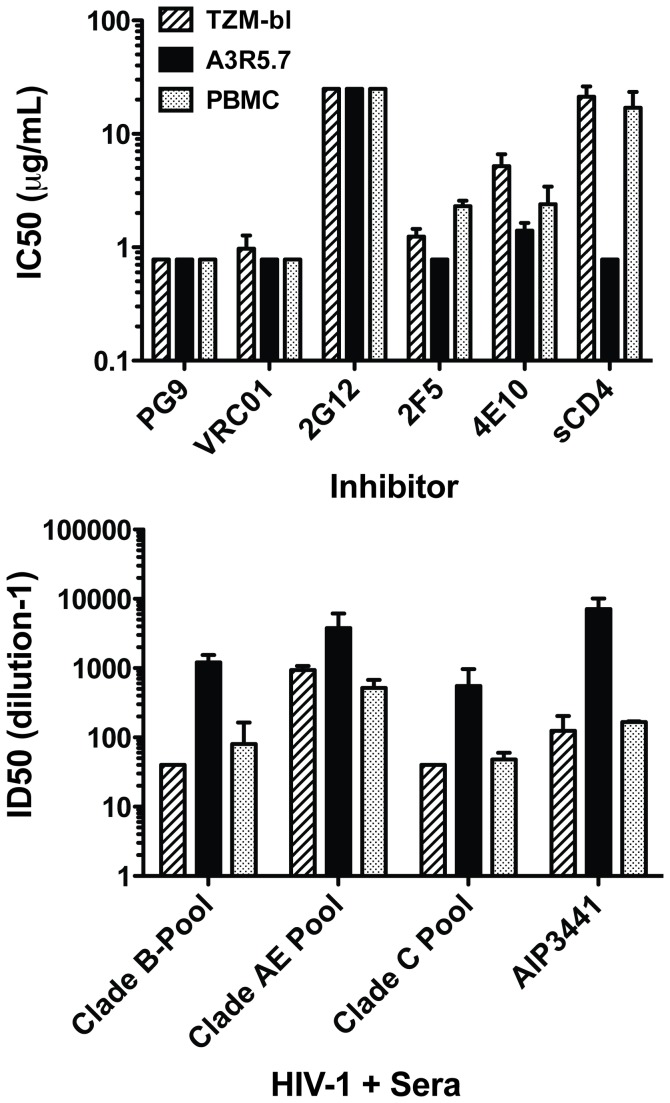
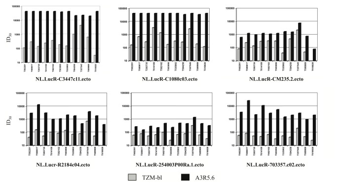
Sensitive assays are needed to meaningfully assess low levels of neutralizing antibodies (NAbs) that may be important for protection against the acquisition of HIV-1 infection in vaccine recipients. The current assay of choice uses a non-lymphoid cell line (TZM-bl) that may lack sensitivity owing to over expression of CD4 and CCR5. We used transfection of a human CD4+/CXCR4+/α4β7+ T-lymphoblastoid cell line (A3.01) with a CMV IE promoter-driven CCR5neo vector to stably express CCR5. The resulting line, designated A3R5, is permissive to a wide range of CCR5-tropic circulating strains of HIV-1, including HIV-1 molecular clones containing a Tat-inducible Renilla luciferase reporter gene and expressing multiple Env subtypes. Flow cytometric analysis found CCR5 surface expression on A3R5 cells to be markedly less than TZM-bl but similar to CD3.8 stimulated PBMC. More importantly, neutralization mediated by a diverse panel of monoclonal antibodies, HIV-1 positive polyclonal sera and sCD4 was consistently greater in A3R5 compared to TZM-bl cells. The A3R5 cell line provides a novel approach to guide the development and qualification of promising new HIV-1 vaccine immunogens.


Conflict of interest statement

Competing Interests: The authors have declared that no competing interests exist.

Figures

Figure 1
Figure 1. Initial characterization of A3R5 cell lines.
A. CCR5 surface receptor number in sequentially sorted A3R5 cell lines. For each sort, 2.0 x 10⁷ cells were subjected to fluorescence activated cell sorting with the brightest population (top 25%) selected for expansion. Receptor number was calculated using Simply Cellular Beads. In general, each successive sort resulted in increasing CCR5 intensity on the cell surface. B. Titration of NL.LucR-env.ecto in A3R5.6, TZM-bl and PHA/IL2 PBMC. Replication competent reporter viruses encoding subtype B env (NL-LucR.T2A-SF162.ecto, NL-LucR.T2A-BaL.ecto) and subtype AE env (NL-LucR.T2A-CM235.ecto) were titered in each target cell. Background luciferase activity (cell control wells) was subtracted prior to plotting. The data show similar virus growth in the three target cells. C. Infectivity of IMC.LucR viruses from multiple subtypes in various CCR5-expressing cell lines. IMC.LucR from subtypes B, A/E and C were screened at a 1:3 dilution in triplicate for their ability to infect four different sorts of A3R5 cells as well as the SupT1.CCR5 and CEM.NKR.CCR5.Luc cell lines. The average relative light unit (RLU) values are indicated. IMC described in Table S1. While all cell lines supported IMC.LucR replication, infection was determined to be optimal in the A3R5.7 cell line. D. Titration of IMC.LucR in A3R5.6 and A3R5.7 cells. Replication competent reporter viruses encoding subtype B Env IMC (NL.LucR-SF162.ecto and NL.LucR-BaL.ecto) and subtype CRF_01 AE Env IMC (CM235, C1080, 0503M02138 and R2184 with backbone: NL.LucR-env.ecto) and subtype C Env IMC (ETH2220.LucR-ETH2220) were titered in the presence of DEAE-Dextran. Virus replication was measured as relative luminescence units (RLUs). Background luciferase activity (cell control wells) was similar in both cell lines and subtracted prior to plotting. Data represents the mean of three replicates with standard error bars (SEM). The data indicate no significant difference in IMC.LucR growth between the A3R5.6 and A3R5.7 cell lines (paired t-test;p0.5).
Figure 2
Figure 2. Receptor expression in A3R5.7 cells.
A. Flow cytometric analysis of CD4, CCR5 and α4β7 expression in the A3R5.7 cell line. 0.5 x 10⁶ cells were singly stained for 30 minutes with fluorochrome-conjugated antibodies as shown followed by fixation in 2% paraformaldehyde. Data are representative of at least two independent experiments. Isotype controls are shown in grey. Nearly all cells were positive for CD4 and CCR5 while approximately half were positive for α4β7. B. Comparison of cell surface CD4, CCR5 and α4β7 receptor densities in various cell targets. 0.5 x 10⁶ cells were stained with fluorochrome-conjugated antibodies and compared to defined populations of similarly stained Quantum Simply Cellular beads. PBMC were stimulated with CD3.8 bi-specific antibody in the presence of 50U/mL rhIL-2. Assuming monovalent antibody-to-surface receptor binding, the Antibody Binding Capacity (ABC) calculated is equivalent to receptors/cell. Data represents the mean of two separate experiments. TZM-bl cells express high levels of CD4 and CCR5 but are negative for α4β7 while A3R5.7 cells possess CCR5 and α4β7 densities more similar to PBMC. CD4 expression on TZM-bl was beyond assay range.
Figure 3
Figure 3. Subtype B neutralization in A3R5 cells and TZM-bl.
A-C. Neutralization sensitivity of three tier 2 subtype B IMC.LucR transmitted/founder viruses to sCD4 and a panel of four mAbs in two A3R5 cell lines and TZM-bl. With the exception where no inhibition was noted for any cell line, the A3R5 cell lines showed greater neutralization for all inhibitors tested compared to TZM-bl. D. Grouped inhibitor comparison between cell lines. When grouped by cell line using the non-parametric Mann-Whitney test, there was a significant difference between the A3R5 cell lines and TZM-bl (Mann-Whitney test; p<0.001). IC50 titers are the concentration of inhibitor at which the Renilla luciferase signal (RLUs) was reduced by 50% compared to the virus control (no inhibitor). Inhibitor concentrations ranged from 0.78 μg/mL to 25 μg/mL. All assays were performed in the presence of DEAE-Dextran.
Figure 4
Figure 4. Neutralization of a tier 2 CRF_01 AE IMC.LucR virus in A3R5.7, TZM-bl and CD3.8 bi-specific antibody stimulated PBMC cell targets.
A. Neutralization with epitope-specific inhibitors. NL.LucR-CM235.ecto was tested against a panel of monoclonal antibodies and sCD4 in each cell line. The highly potent PG9 ad VRC01 mAbs demonstrated IC50 titers below the limits of the assay (<0.78μg/mL). This chronic envelope IMC was insensitive to 2G12 (>25μg/mL) in all target cells. NL.LucR-CM235.ecto was more sensitive to the inhibitors 2F5, 4E10 and sCD4 in the A3R5.7 target cell compared to both TZM-bl and PBMC, reaching significance with sCD4 (Unpaired t-test with Welch correction; p<0.05). Inhibitors ranged from 0.78μg/mL to 25 μg/mL. B. Neutralization with polyclonal sera. NL.LucR-CM235.ecto was tested against a panel of pooled HIV-1 “+” sera/plasma and the individual AE-specific sera AIP3441 in each cell line. Sera dilutions ranged from 40-40,960. All sera appeared more potent in the A3R5.7 cell line compared to either TZM-bl or PBMC.
Figure 5
Figure 5. Comparison of neutralization sensitivity in A3R5 and TZM-bl cell lines.
A panel of eleven HIV+ CRF_01 AE sera was assayed against two tier 1 (C3347.c11 and C1080.c03) and four tier 2 (CM235-2, R2184.c04, 254003P00Ra.1 and 703357.c02) CRR_01 AE Env.IMC.LucR viruses in the A3R5.6 and TZM-bl cell lines. ID50 titers are the dilution of serum at which the Renilla luciferase signal was reduced by 50% compared to the virus control (no serum). Neutralization sensitivity was significantly greater (p<0.001; Wilcoxon Rank Sum) in the A3R5 cell line compared to TZM-bl for all virus/sera pairs tested. Similar results were obtained with the A3R5.7 cell line.

References

    1. Rerks-Ngarm S, Pitisuttithum P, Nitayaphan S, Kaewkungwal J, Chiu J et al. (2009) Vaccination with ALVAC and AIDSVAX to prevent HIV-1 infection in Thailand. N Engl J Med 361: 2209-2220. doi:10.1056/NEJMoa0908492. PubMed: 19843557. - DOI - PubMed
    1. Montefiori DC, Karnasuta C, Huang Y, Ahmed H, Gilbert P et al. (2012) Magnitude and breadth of the neutralizing antibody response in the RV144 and Vax003 HIV-1 vaccine efficacy trials. J Infect Dis 206: 431-441. doi:10.1093/infdis/jis367. PubMed: 22634875. - DOI - PMC - PubMed
    1. Nitayaphan S, Pitisuttithum P, Karnasuta C, Eamsila C, de Souza M et al. (2004) Safety and immunogenicity of an HIV subtype B and E prime-boost vaccine combination in HIV-negative Thai adults. J Infect Dis 190: 702-706. doi:10.1086/422258. PubMed: 15272397. - DOI - PubMed
    1. Haynes BF, Gilbert PB, McElrath MJ, Zolla-Pazner S, Tomaras GD et al. (2012) Immune-correlates analysis of an HIV-1 vaccine efficacy trial. N Engl J Med 366: 1275-1286. doi:10.1056/NEJMoa1113425. PubMed: 22475592. - DOI - PMC - PubMed
    1. Mascola JR, Montefiori DC (2010) The role of antibodies in HIV vaccines. Annu Rev Immunol 28: 413-444. doi:10.1146/annurev-immunol-030409-101256. PubMed: 20192810. - DOI - PubMed

Publication types