. 2013 Nov 29;8(11):e79541.
doi: 10.1371/journal.pone.0079541. eCollection 2013.

Use of the γ-H2AX assay to investigate DNA repair dynamics following multiple radiation exposures

Affiliations

Use of the γ-H2AX assay to investigate DNA repair dynamics following multiple radiation exposures

Luca G Mariotti et al. PLoS One. .

Abstract

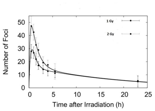
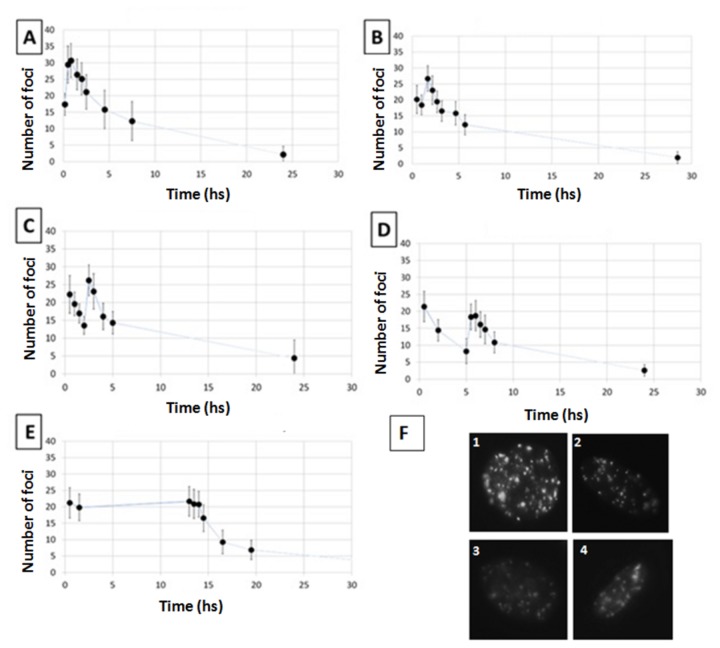
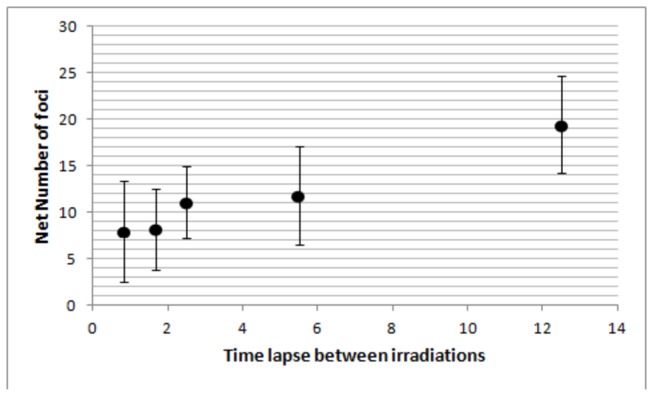
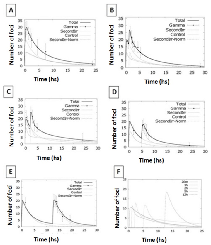
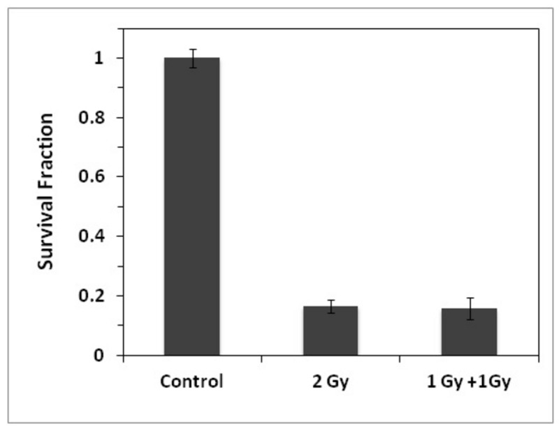
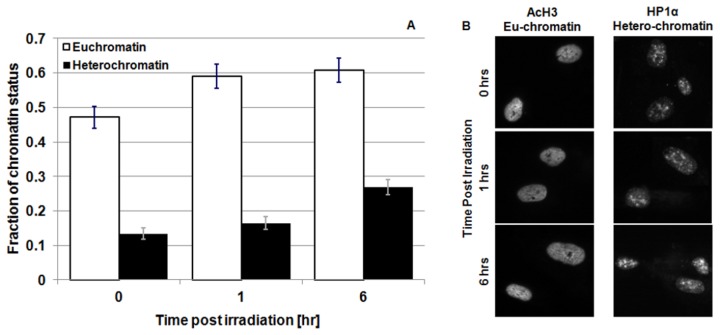
Radiation therapy is one of the most common and effective strategies used to treat cancer. The irradiation is usually performed with a fractionated scheme, where the dose required to kill tumour cells is given in several sessions, spaced by specific time intervals, to allow healthy tissue recovery. In this work, we examined the DNA repair dynamics of cells exposed to radiation delivered in fractions, by assessing the response of histone-2AX (H2AX) phosphorylation (γ-H2AX), a marker of DNA double strand breaks. γ-H2AX foci induction and disappearance were monitored following split dose irradiation experiments in which time interval between exposure and dose were varied. Experimental data have been coupled to an analytical theoretical model, in order to quantify key parameters involved in the foci induction process. Induction of γ-H2AX foci was found to be affected by the initial radiation exposure with a smaller number of foci induced by subsequent exposures. This was compared to chromatin relaxation and cell survival. The time needed for full recovery of γ-H2AX foci induction was quantified (12 hours) and the 1:1 relationship between radiation induced DNA double strand breaks and foci numbers was critically assessed in the multiple irradiation scenarios.


Conflict of interest statement

Competing Interests: The authors have declared that no competing interests exist.

Figures

Figure 1
Figure 1. Foci kinetics following an acute irradiation with 225 kVp X-rays.
Panel A. Experimental data and fit (from eq. 3) of γ-H2AX foci after exposure to 1 Gy (full circles) and 2 Gy (open squares) for 225 kVp X-rays. Error bars represent one standard error of the mean of 3 independent experiments. Panel B. Representative pictures of γ-H2AX foci after irradiation with 1 and 2 Gy of 225 kVp X-rays. Upper figures represent 2 examples of a small field of view, whereas the middle and lower pictures show view at a single cell level.
Figure 2
Figure 2. Foci kinetics following an acute irradiation with 30 kVp X-rays.
Experimental data and fit (from eq. 1) of γ-H2AX foci after exposure to 1 Gy (full circles) and 2 Gy (dotted circles) for 30 kVp X-rays. Error bars represent one standard error of the mean of 3 independent experiments.
Figure 3
Figure 3. γ-H2AX foci kinetics following split multiple radiation exposures.
Number of γ-H2AX foci per cell after exposure to 1 Gy at time 0 plus 1 Gy delivered20 min later (panel A), 1 hour later (panel B), 2 hours later (panel C), 5 hours later (panel D) and 12 hours later (panel E) using 225 kVp X-rays. In Panel F representative pictures of cells in different exposures scenario are presented, i.e. cells fixed after 30 minutes in the cases of 20 minutes split dose exposure (Figure 1), 1 hour split dose exposure (Figure 2), 2 hours split dose exposure (Figure 3) and 5 hours split dose exposure (Figure 4). Error bars represent one standard error of the mean of 3 independent experiments. Lines are guides for the eyes.
Figure 4
Figure 4. Net number of γ-H2AX foci induced by the second radiation exposure.
Estimated number of γ-H2AX foci induced by the second irradiation only as calculated by subtracting the predicted residual number of foci from the first exposure (using the single irradiation kinetic data) from the total number of foci measured 30 minutes after the second irradiation.
Figure 5
Figure 5. Fitting kinetics for split dose irradiation exposures.
Number of γ-H2AX foci per cell after exposure to 1 Gy at time 0 plus 1 Gy delivered 20 minutes, 1 hr, 2 hr, 5 hr or 12 hr later using 225 kVp X-rays. Solid line represents the modelling function for the split dose scenario. Dotted lines represent the modelling function for the individual radiation exposures. Kinetics for the second exposure only are reported in the last panel.
Figure 6
Figure 6. Clonogenic data survival following single and split dose irradiations.
Comparison of clonogenic survival following acute (2 Gy) and split dose (1 Gy + 1 Gy) exposures with 1 hr time gap. Error bars represent the standard error of the mean of 3 independent experiments.
Figure 7
Figure 7. Eu/hetero-chromatin change following radiation exposure.
Panel A: Amount of eu- (acH3 staining - light bars) and hetero-chromatin (HP1α. staining - dark bars) in cell nuclei as a function of time after 1 Gy of 225 kVp X-rays irradiation of 3 independent experiments. The analysis of the experimental data has been performed with a symmetric T-Student test. Panel B: Representative picture of eu- and hetero-chromatin staining .
Figure 8
Figure 8. Split dose irradiations with small initial dose.
Number of γ-H2AX foci after exposure to 0.1 Gy at time 0 plus 1 Gy delivered 1 hr after (left column) and 4 hr later (right column). Top panels report the experimental data;bottom panels, data are fitted using the modelling equation (4). In the bottom panels, the solid line represents the modelling function for the split dose scenario. The dotted lines represent the modelling function for the individual radiation exposures. Error bars represent the standard error of the mean of 3 independent experiments.

References

    1. Kavanagh J, Redmond K, Schettino G, Prise K (2013) DSB Repair - A radiation perspective. Antioxid Redox Signal [Epub ahead of print]. - PubMed
    1. Alloni D, Campa A, Friedland W, Mariotti L, Ottolenghi A (2012) Track structure, radiation quality and initial radiobiological events: Considerations based on the PARTRAC code experience. Int J Radiat Biol 88: 77–86. doi:10.3109/09553002.2011.627976. PubMed: 21957961. - DOI - PubMed
    1. Alloni D, Campa A, Friedland W, Mariotti L, Ottolenghi A (2013) Integration of Monte Carlo Simulations with PFGE Experimental Data Yields Constant RBE of 2.3 for DNA Double-Strand Break Induction by Nitrogen Ions between 125 and 225 keV/μm LET. Radiat Res 179: 690-697. doi:10.1667/R3043.1. PubMed: 23647004. - DOI - PubMed
    1. Alloni D, Campa A, Belli M, Esposito G, Mariotti L et al. (2011) Monte Carlo evaluation of DNA fragmentation spectra induced by different radiation qualities. Radiat Protect Dosim 143: 226-231. doi:10.1093/rpd/ncq384. PubMed: 21084331. - DOI - PubMed
    1. Khanna KK, Jackson SP (2001) DNA double-strand breaks: signaling, repair and the cancer connection. Nat Genet 27: 247-254. doi:10.1038/85798. PubMed: 11242102. - DOI - PubMed

Publication types