. 2013 Nov 28;8(11):e80995.
doi: 10.1371/journal.pone.0080995. eCollection 2013.

Molecular markers for granulovacuolar degeneration are present in rimmed vacuoles

Affiliations

Molecular markers for granulovacuolar degeneration are present in rimmed vacuoles

Masahiro Nakamori et al. PLoS One. .

Expression of concern in

Abstract

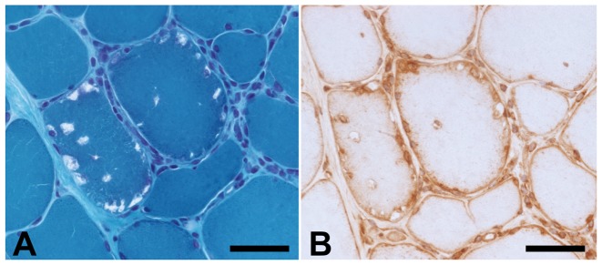
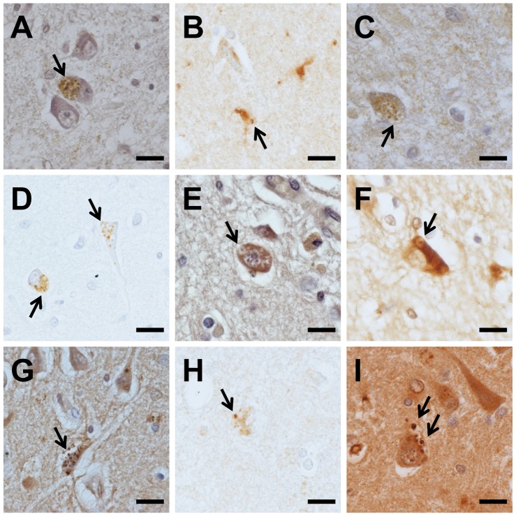
Background: Rimmed vacuoles (RVs) are round-oval cytoplasmic inclusions, detected in muscle cells of patients with myopathies, such as inclusion body myositis (IBM) and distal myopathy with RVs (DMRV). Granulovacuolar degeneration (GVD) bodies are spherical vacuoles containing argentophilic and hematoxyphilic granules, and are one of the pathological hallmarks commonly found in hippocampal pyramidal neurons of patients with aging-related neurodegenerative diseases, such as Alzheimer's disease and Parkinson's disease. These diseases are common in the elderly and share some pathological features. Therefore, we hypothesized that mechanisms of vacuolar formation in RVs and GVD bodies are common despite their role in two differing pathologies. We explored the components of RVs by immunohistochemistry, using antibodies for GVD markers.

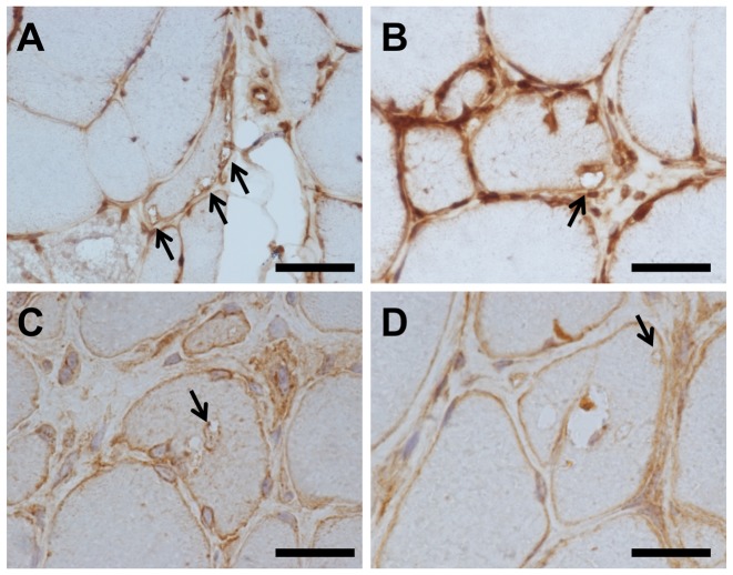
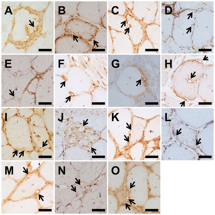
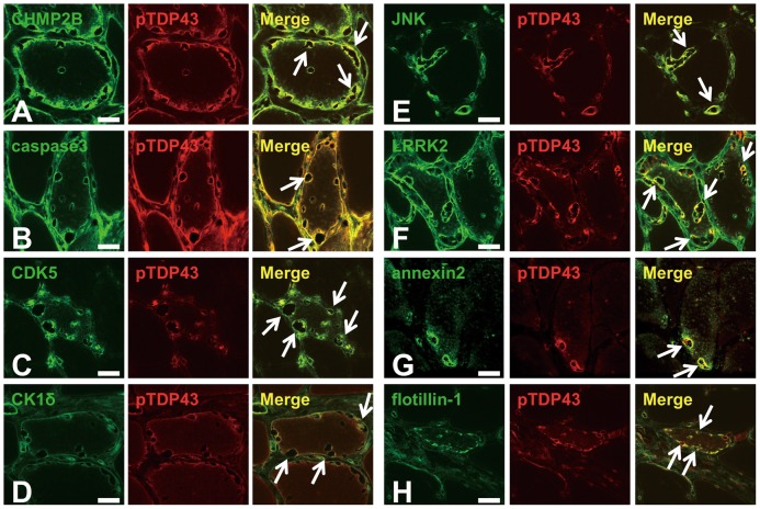
Methods: Subjects included one AD case, eight cases of sporadic IBM, and three cases of DMRV. We compared immunoreactivity and staining patterns for GVD markers. These markers included: (1) tau-modifying proteins (caspase 3, cyclin-dependent kinase 5 [CDK5], casein kinase 1δ [CK1δ], and c-jun N-terminal kinase [JNK]), (2) lipid raft-associated materials (annexin 2, leucine-rich repeat kinase 2 [LRRK2], and flotillin-1), and (3) other markers (charged multi-vesicular body protein 2B [CHMP2B] and phosphorylated transactive response DNA binding protein-43 [pTDP43]) in both GVD bodies and RVs. Furthermore, we performed double staining of each GVD marker with pTDP43 to verify the co-localization.

Results: GVD markers, including lipid raft-associated proteins and tau kinases, were detected in RVs. CHMP2B, pTDP43, caspase 3, LRRK2, annexin 2 and flotillin-1 were detected on the rim and were diffusely distributed in the cytoplasm of RV-positive fibers. CDK5, CK1δ and JNK were detected only on the rim. In double staining experiments, all GVD markers colocalized with pTDP43 in RVs.

Conclusions: These results suggest that RVs of muscle cells and GVD bodies of neurons share a number of molecules, such as raft-related proteins and tau-modifying proteins.


Conflict of interest statement

Competing Interests: The authors have declared that no competing interests exist.

Figures

Figure 1
Figure 1. CHMP2B-positive structures corresponding to RVs.
Modified Gomori trichrome staining, and immunohistochemical staining of serial sections using anti-charged multivesicular body protein 2B (CHMP2B) antibody, in an sporadic inclusion body myositis (s-IBM) case (case 1). Rimmed vacuoles (RVs) identified by modified Gomori trichrome staining (A) and the same structure immunopositive for CHMP2B (B). Scale bars  = 20 µm.
Figure 2
Figure 2. Immunohistochemistry for CHMP2B in muscle fibers of s-IBM and DMRV cases.
RVs in all s-IBM (A–C) and distal myopathy with RVs (DMRV) (D) cases detected by anti- CHMP2B antibody. Arrows indicate RVs. Scale bar  = 20 µm.
Figure 3
Figure 3. Immunohistochemistry for GVD markers in the Alzheimer's disease hippocampus.
Anti-CHMP2B (A), anti-caspase3 (B), anti-cyclin-dependent kinase 5 (CDK5) (C), anti-casein kinase 1δ (CK1δ) (D), anti-c-jun N-terminal kinase (JNK) (E), anti-leucine-rich repeat kinase 2 (LRRK2) (F), anti-annexin2 (G), anti-flotillin-1 (H), and anti-phosphorylated transactive response DNA binding protein-43 (pTDP43) (I). Arrows indicate granulovacuolar degeneration (GVD) bodies. Scale bars  = 20 µm.
Figure 4
Figure 4. Immunohistochemistry for GVD markers in muscle fibers of s-IBM cases.
Anti-CHMP2B (A), anti-caspase3 (B, C), anti-CDK5 (D), anti- CK1δ (E, F), anti-JNK (G, H), anti-LRRK2 (I), anti-annexin2 (J, K), anti-flotillin-1 (L, M), and anti-pTDP43 (N, O). (B, C), (E, F), (G,H), (J, K), (L, M) and (N, O) indicate 2 independent antibodies for the identification of each protein. Arrows indicate RVs. Scale bar  = 20 µm.
Figure 5
Figure 5. Representative confocal laser scanning micrograph.
Double immunofluorescence labeling and merged images in muscle sections from patients with s-IBM for CHMP2B (green) and pTDP43 (red) (A), caspase3 (green) and pTDP43 (red) (B), CDK5 (green) and pTDP43 (red) (C), CK1δ (green) and pTDP43 (red) (D), JNK (green) and pTDP43 (red) (E), LRRK2 (green) and pTDP43 (red) (F), annexin 2 (green) and pTDP43 (red) (G), and flotillin-1 (green) and pTDP43 (red) (H). Arrows indicate RVs. Scale bar  = 20 µm.

References

    1. Fukuhara N, Kumamoto T, Tsubaki T (1980) Rimmed vacuoles. Acta Neuropathol 51: 229–235. - PubMed
    1. Momma K, Noguchi S, Malicdan MC, Hayashi YK, Minami N, et al. (2012) Rimmed vacuoles in Becker muscular dystrophy have similar features with inclusion myopathies. PLoS One 7: e52002. - PMC - PubMed
    1. Bosch EP, Gowans JD, Munsat T (1979) Inflammatory myopathy in oculopharyngeal dystrophy. Muscle Nerve 2: 73–77. - PubMed
    1. Askanas V, Engel WK (2008) Inclusion-body myositis: muscle-fiber molecular pathology and possible pathogenic significance of its similarity to Alzheimer's and Parkinson's disease brains. Acta Neuropathol 116: 583–595. - PMC - PubMed
    1. Askanas V, Engel WK, Nogalska A (2009) Inclusion body myositis: a degenerative muscle disease associated with intra-muscle fiber multi-protein aggregates, proteasome inhibition, endoplasmic reticulum stress and decreased lysosomal degradation. Brain Pathol 19: 493–506. - PMC - PubMed

Publication types

Substances