. 2013 Nov 28;8(11):e81184.
doi: 10.1371/journal.pone.0081184. eCollection 2013.

Natural stilbenoids isolated from grapevine exhibiting inhibitory effects against HIV-1 integrase and eukaryote MOS1 transposase in vitro activities

Affiliations

Natural stilbenoids isolated from grapevine exhibiting inhibitory effects against HIV-1 integrase and eukaryote MOS1 transposase in vitro activities

Aude Pflieger et al. PLoS One. .

Abstract

Polynucleotidyl transferases are enzymes involved in several DNA mobility mechanisms in prokaryotes and eukaryotes. Some of them such as retroviral integrases are crucial for pathogenous processes and are therefore good candidates for therapeutic approaches. To identify new therapeutic compounds and new tools for investigating the common functional features of these proteins, we addressed the inhibition properties of natural stilbenoids deriving from resveratrol on two models: the HIV-1 integrase and the eukaryote MOS-1 transposase. Two resveratrol dimers, leachianol F and G, were isolated for the first time in Vitis along with fourteen known stilbenoids: E-resveratrol, E-piceid, E-pterostilbene, E-piceatannol, (+)-E-ε-viniferin, E-ε-viniferinglucoside, E-scirpusin A, quadragularin A, ampelopsin A, pallidol, E-miyabenol C, E-vitisin B, hopeaphenol, and isohopeaphenol and were purified from stalks of Vitis vinifera (Vitaceae), and moracin M from stem bark of Milliciaexelsa (Moraceae). These compounds were tested in in vitro and in vivo assays reproducing the activity of both enzymes. Several molecules presented significant inhibition on both systems. Some of the molecules were found to be active against both proteins while others were specific for one of the two models. Comparison of the differential effects of the molecules suggested that the compounds could target specific intermediate nucleocomplexes of the reactions. Additionally E-pterostilbene was found active on the early lentiviral replication steps in lentiviruses transduced cells. Consequently, in addition to representing new original lead compounds for further modelling of new active agents against HIV-1 integrase, these molecules could be good tools for identifying such reaction intermediates in DNA mobility processes.


Conflict of interest statement

Competing Interests: The authors have declared that no competing interests exist.

Figures

Figure 1
Figure 1. General structure of resveratrol derivative stilbenes purified form Vitis vinifera grapevine.
The compounds are divided into three families: monomers, dimers and higher order oligomers of the resveratrol motif. A list of the acronyms used in this work is reported in the figure.
Figure 2
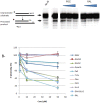
Figure 2. Selection of stilbenes inhibiting in vitro HIV-1 integration.
Concerted integration assay was performed without IN or using 600 µM of compounds. The reaction products were loaded onto 1% agarose gel. The position and the structure of the different products obtained after half-site (HSI), full-site (FSI) and donor/donor integration (d/d) are shown in A as well as the integration profile obtained in presence of the DMSO control, selected (here DIM1D) or unselected molecule (here TRI1). The integration products were quantified as reported in materials and methods and the integration inhibition is shown in B using the integration activity detected in presence of DMSO as reference. All quantifications are shown as the means from at least three independent experiments ± standard deviation (error bars). We also reported the inhibitory effect observed with 10 µM raltegravir (RAL).
Figure 3
Figure 3. Effect of stilbene compounds on in vitro HIV-1 concerted integration.
Concerted integration assays were performed using 600(A) or unprocessed (B) donor DNA incubated with increasing concentration of compounds. The integration products were quantified as reported in materials and methods section and the percentage of integration is shown as the means from at least three independent experiments ± standard deviation (error bars). We also reported the inhibitory effect observed with raltegravir (RAL).
Figure 4
Figure 4. Effect of stilbene compounds on in vitro 3′processing reaction catalysed by HIV-1 integrase.
Standard 3′processing assay was performed as reported in materials and methods section using 600 nM IN, 1 pmol of 5′ radiolabelled 21 bp ODN 70/72 mimicking the viral U5 end and increasing concentration of molecules. An example of 3′ processing profile obtained with a selected inhibitory compound (here E-piceatannol (PICE) and the raltegravir (RAL) control molecule is shown in A as well as the position and the structure of the expected 19 bp reaction product. The 19 bp processed product was quantified as reported in materials and methods section and the percentage of 3′ processing is shown in B as the average from at least three independent experiments ± standard deviation (error bars).
Figure 5
Figure 5. Effect of selected molecules on the early steps of VSV-G pseudotyped lentiviruses replication in 293T cells.
After 24materials and methods (A). The cell survival is reported as a percentage of control without molecules and as the means from at least three independent experiments ± standard deviation (error bars). The effect on replication was measured by FACS quantification of the GFP expressed by the cells transduced with VSV-G pseudotyped lentiviruses (B). Results are reported as a percentage of GFP positive cells as the average from at least three independent experiments ± standard deviation. Total (C), Integrated, 2LTR (D) and 1LTR (E) circles viral DNA were quantified (8 h post-infection of the total DNA and 24 h post-infection for the integrated and 2LTR circle DNA) by quantitative PCR. An example of quantification obtained with an inhibitory compound (here Pter) is shown. Total viral DNA is reported as percentage of control without drug. Integrated and 2LTR circles DNA are reported as a percentage of each form comparing to total viral DNA. Results are reported as the average from at least three independent experiments ± standard deviation, *p<0.05.
Figure 6
Figure 6. Selection by in vitro transposition of natural stilbene compounds active against Mos1 transposase.
(A) in vitro transposition test principle. pBC-3T3 was used both as the donor of pseudo-Mos1 and as the target for integration. It contained the pBR322 tetracycline resistance gene (without promoter) flanked by two Mos1 3′ITR. This plasmid was unable to confer resistance to Escherichia coli cells in tetracycline concentrations over 10 µg/ml. Upon transposition, MOS1 excises the pseudo-Mos1 from one pBC-3T3 molecule, and then triggers its reinsertion within the cat gene of another pBC-3T3 molecule . Transposition events are revealed by promoter tagging, the tetracycline resistance being activated through the cat gene promoter. Transposition rates were quantified as reported in materials and methods section and the transposition inhibition is shown in (B) using the transposition rate obtained in presence of DMSO as reference. Values are the means from at least two independent experiments ± standard deviation (error bars). Only compounds giving a transposition rate inhibition over 50% at 25 µM were assayed at 10 µM.
Figure 7
Figure 7. Effect of the selected molecules on Mos1 excision.
(A) In vitro excision assays were performed using a super coiled plasmid pBC-3T3 and purified MBP-MOS1. The resulting products were loaded onto agarose gel. Data obtained with two molecules (25 µM of DIM1F or HOPE) or DMSO (control) are shown. The pBC-3T3 plasmid used as the substrate is shown in lane2.Molecular weight markers are indicated on the left. The various products are depicted on the right and their positions on the gel are indicated. SC: super coiled donor. First strand nicking at one transposon end generates an open circular product (OC). Second strand nicking linearizes the donor (L), yielding the single-end break product. A similar sequence of nicks at the other transposon end yields the double-end break products, which consist of the plasmid backbone (B) plus the excised transposon fragment (not shown). (B) DNA products detected in the gel were quantified, and the appearance of the backbone (B) was used as a measure of excision. The control assay (DMSO) was taken as the reference (100% excision) and data obtained for each molecule (at 25 and 10 µM) are expressed as excision relative percentage. Each point was repeated at least twice. (C) IC50 were determined for three molecules (Pall, HOPE, and DIM1F) using a three log range of compound concentrations. The percentage inhibition of excision was plotted as a function of the log of compound concentrations. Experimental data were fitted on a sigmoid dose-response curve using Prism software.
Figure 8
Figure 8. Effect of molecules on Mos1 ITR integration.
(A) In vitro integration assays were performed using labelled PC-ITR and purified MBP-MOS1. pBC was used as the target. The resulting products were loaded onto agarose gel and detected using a Storm. Data obtained with two molecules (25 µM of DIM1F or HOPE) or DMSO (control) are shown. The various products are indicated on the right. Each point was repeated at least twice. (B) DNA products detected in the gel were quantified and the appearance of labelled plasmid (integration products) was used as a measure of integration. The control assay (DMSO) was taken as the reference (100% integration) and data obtained for each molecule (at 25 and 10 µM) are shown as relative percentage of integration. (C) IC50 were determined for two molecules (HOPE, and DIM1F) using a four log range of compound concentrations. The percentage inhibition of integration was plotted as a function of the log of compound concentrations. Experimental data were fitted on a sigmoid dose-response curve using Prism software.

References

    1. Engelman A, Mizuuchi K, Craigie R (1991) HIV-1 DNA Integration: Mechanism of viral DNA Cleavage and strand transfer. Cell 67: 1211–1221. - PubMed
    1. Carpentier G, Jaillet J, Pflieger A, Adet J, Renault S, et al. (2011) Transposase-transposase interactions in MOS1 complexes: a biochemical approach. J Mol Biol 405: 892–908. - PubMed
    1. Jaillet J, Genty M, Cambefort J, Rouault JD, Auge-Gouillou C (2012) Regulation of mariner transposition: the peculiar case of Mos1. PLoS One 7: e43365. - PMC - PubMed
    1. Richardson JM, Colloms SD, Finnegan DJ, Walkinshaw MD (2009) Molecular architecture of the Mos1 paired-end complex: the structural basis of DNA transposition in a eukaryote. Cell 138: 1096–1108. - PMC - PubMed
    1. Hare S, Vos AM, Clayton RF, Thuring JW, Cummings MD, et al. (2010) Molecular mechanisms of retroviral integrase inhibition and the evolution of viral resistance. Proc Natl Acad Sci U S A 107: 20057–20062. - PMC - PubMed

Publication types

MeSH terms