. 2013 Nov 29;8(11):e81710.
doi: 10.1371/journal.pone.0081710. eCollection 2013.

Mutational analysis of sclerostin shows importance of the flexible loop and the cystine-knot for Wnt-signaling inhibition

Affiliations

Mutational analysis of sclerostin shows importance of the flexible loop and the cystine-knot for Wnt-signaling inhibition

Verena Boschert et al. PLoS One. .

Erratum in

  • PLoS One. 2014;9(1). doi:10.1371/annotation/067f1197-3290-4d1e-8f77-4d3996a6b9e8

Abstract

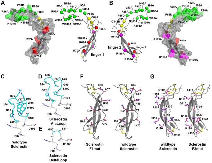
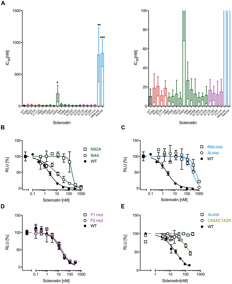
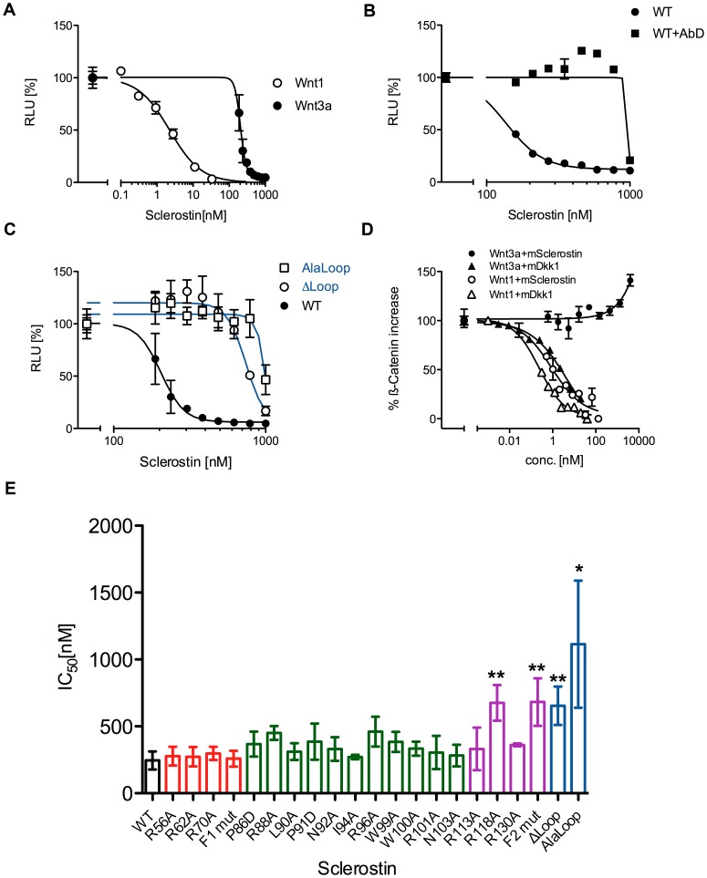
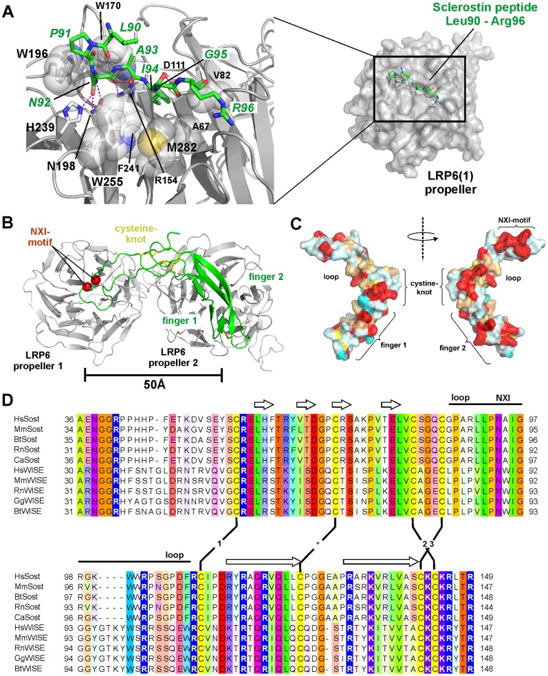
The cystine-knot containing protein Sclerostin is an important negative regulator of bone growth and therefore represents a promising therapeutic target. It exerts its biological task by inhibiting the Wnt (wingless and int1) signaling pathway, which participates in bone formation by promoting the differentiation of mesenchymal stem cells to osteoblasts. The core structure of Sclerostin consists of three loops with the first and third loop (Finger 1 and Finger 2) forming a structured β-sheet and the second loop being unstructured and highly flexible. Biochemical data showed that the flexible loop is important for binding of Sclerostin to Wnt co-receptors of the low-density lipoprotein related-protein family (LRP), by interacting with the Wnt co-receptors LRP5 or -6 it inhibits Wnt signaling. To further examine the structural requirements for Wnt inhibition, we performed an extensive mutational study within all three loops of the Sclerostin core domain involving single and multiple mutations as well as truncation of important regions. By this approach we could confirm the importance of the second loop and especially of amino acids Asn92 and Ile94 for binding to LRP6. Based on a Sclerostin variant found in a Turkish family suffering from Sclerosteosis we generated a Sclerostin mutant with cysteines 84 and 142 exchanged thereby removing the third disulfide bond of the cystine-knot. This mutant binds to LRP6 with reduced binding affinity and also exhibits a strongly reduced inhibitory activity against Wnt1 thereby showing that also elements outside the flexible loop are important for inhibition of Wnt by Sclerostin. Additionally, we examined the effect of the mutations on the inhibition of two different Wnt proteins, Wnt3a and Wnt1. We could detect clear differences in the inhibition of these proteins, suggesting that the mechanism by which Sclerostin antagonizes Wnt1 and Wnt3a is fundamentally different.


Conflict of interest statement

Competing Interests: The authors have declared that no competing interests exist.

Figures

Figure 1
Figure 1. Overview of investigated Sclerostin variants.
(A) Surface and secondary structure representation of Sclerostin with residues selected for single point mutations shown as spheres. Residues located in the flexible loop of Sclerostin are colored green, amino acid residues in finger 1 and 2 are indicated in red and magenta, cysteine residues are shown in yellow, respectively. (B) As in (A) but rotated around the y-axis by 180°. (C) Detailed view of the loop region of Sclerostin comprising the residues Gly85 to Asp106. The backbone atoms are shown as ribbon, side chains of non-glycine residues as ball-and-stick models. The region consisting of Leu90 to Asn103 used in the mutational analysis is marked with carbon atoms colored in cyan. (D) Loop region of the Sclerostin multivariant Alaloop. (E) Loop region of the Sclerostin truncation variant ΔLoop, residues Leu90 to Asn103 were replaced by a glycine-serine linker. (F) View of the finger region of wildtype Sclerostin and the Sclerostin multivariant F1mut. Residues shown as ball-and-stick models (carbon atoms colored in red) in finger 1 of wildtype Sclerostin were exchanged to those shown on the left panel. (G) Residues in finger 2 of Sclerostin and their counterpart in the Sclerostin multivariant F2mut are indicated as ball-and-stick models with the carbon atoms colored in magenta.
Figure 2
Figure 2. Reporter gene assay to measure the neutralization of mWnt1 activity by wildtype and Sclerostin variants.
HEK293TSA cells stably transfected with the Wnt-responsive luciferase reporter construct SuperTOPFlash were transfected with an expression construct for murine Wnt1. After 48 h the cells were stimulated with serial dilutions of wildtype (WT) Sclerostin or variants thereof. (A) Overview of the efficiency of the Sclerostin proteins to neutralize Wnt1 driven luciferase expression (IC50 values are shown as bar diagram). The right panel shows a magnification of the data shown in the left panel. Data represents means with standard deviations (SD) of at least three independent experiments. *: P<0.05, **: P<0.01, ***: P<0.001 (student's t-test with data obtained for WT Sclerostin). (B–D) Measurements showing the dose-dependency of selected mutant Sclerostin proteins. IC50 values of these experiments are included in the overview shown in (A). Measurements were done in duplicate. (E) Reporter gene assay using supernatants of HEK Freestyle cells expressing Sclerostin mutants C84AC142R, ΔLoop or wildtype Sclerostin. Data points represent duplicates. To highlight the location of the mutation in the Sclerostin structure the same color-coding as in Figure 1 is used.
Figure 4
Figure 4. Analysis of binding of wildtype Sclerostin and different variants to hLRP6.
(A) COS-1 cells were either transfected with an expression construct encoding for hLRP6 (COS-1 + hLRP6) or mock-transfected using empty vector (COS1). Cells were then incubated with I125-labeled wildtype Sclerostin (I125). Protein bound to the cell surface was chemically crosslinked in the presence of 2 µg unlabeled wildtype Sclerostin or the indicated Sclerostin variants for competition, corresponding to a 100-fold excess of unlabeled protein. The radioactivity bound to the cells was subsequently analyzed by autoradiography. Shown bands were all obtained in the same experiment. The black bar indicates that the picture was cut to omit samples that are not relevant for this publication. (B) Quantitative autoradiography analysis of several experiments as shown in (A). All bars were normalized by setting the intensity of the protein band obtained from addition of unlabeled wildtype Sclerostin to partially compete off I125-labeled wildtype Sclerostin to 1. Data represent means with standard deviations (SD) of at least three independent experiments. *: P<0.05, **: P<0.01 (student's t-test with data obtained for variant R56A). (C) Western Blot of a pull down experiment with biotinylated Sclerostin mutants C84AC142R, ΔLoop and WT Sclerostin. Sclerostin proteins were incubated with purified human LRP6 protein fragment comprising propeller domains E1 and E2 (LRP6E1E2). For pull down of protein complexes streptavidin-agarose beads were used. Input: Samples before beads were added, beads: Beads after incubation and washing. Proteins were detected using an antibody specific for the His6-tag of the proteins. One representative out of four experiments is shown.
Figure 3
Figure 3. Inhibition of Wnt3a signaling by Sclerostin.
(A) HEK293TSA cells stably transfected with the Wnt-responsible luciferase reporter construct SuperTOPFlash were stimulated with 1.5 nM recombinant murine Wnt3a and serial dilutions of murine Sclerostin. Shown is the resulting dose response curve in comparison to the dose response curve of mWnt1-transfected cells as shown in figure 3. (B) mWnt3a-derived luciferase signal obtained in presence of Sclerostin wildtype (WT) and 2 µM Sclerostin specific antibody AbD09097 (AbD) or with WT alone. (C) Reporter gene assay as depicted in (A) and (B) showing the dose-dependency of Sclerostin Alaloop and ΔLoop in comparison to WT Sclerostin. Measurements were done in duplicate. (D) Measurement of the intracellular level of β-Catenin upon mWnt3a and mWnt1 stimulation in presence of Sclerostin or Dkk1 using In-Cell Western. The fluorescence signal determined from the antibody against β-Catenin was normalized for cell number using DNA staining with DRAQ5. Signal of non-transfected/untreated cells was set to 0% and signal of cells treated/transfected with Wnt proteins alone was set to 100%. A typical experiment out of three is shown; datapoints represent means with SD of two independent measurements. (E) Overview of the efficiency of Sclerostin WT and mutant proteins to neutralize Wnt3a driven luciferase expression (IC50 values represented as bar diagram). Data represent means with standard deviation (SD) of at least two independent experiments. *: P<0.05 **: P<0.01 (student's t-test with data obtained for wildtype Sclerostin). To highlight the location of the mutation in the Sclerostin structure the same color-coding as in figure 2 is used.
Figure 5
Figure 5. Modeling of a Sclerostin-LRP6E1E2 complex indicates additional interactions apart from the NXI motif.
(A) Interaction scheme of a 7 mer peptide representing the loop tip amino acid sequence of Sclerostin (residues Leu90 to Arg96) bound into the cleft of the β-propeller domain 1 of LRP6 (PDB entry 3SOV, [27]). Two main binding determinants were identified in the Sclerostin-derived peptide, Asn92 and Ile94, which are engaged in several polar bonds (Asn92) as well as hydrophobic interactions (Ile94) to facilitate recognition and binding. Residues of LRP6 involved in the interaction with Asn92 and Ile94 are shown in the magnification on the left. (B) Theoretical model of the structured core domain of Sclerostin docked onto the propeller 1-2 fragment of LRP6. The two cavities formed by propeller domain 1 and 2 are separated by 50 Å. The extended architecture of Sclerostin (coincidentally measuring also 50 Å in length) might therefore contact propeller domain 1 and 2 (the latter with parts of Sclerostin finger 1 or 2) possibly explaining the higher binding affinity of Sclerostin to LRP6 fragments containing both propeller domains compared to LRP6 propeller 1 alone. (C) Surface representation of murine Sclerostin (left and right presentation are rotated by 180° around the y-axis) color-coded on the basis of an amino acid sequence alignment (D). The colors mark the level of amino acid identity/homology between Sclerostin and Wise proteins of different organisms with red highlighting invariant residues, orange for exchanges by homologous amino acids, and light and dark blue marking variable residue positions. (D) Sequence alignment of the core domain of Sclerostin and the related Wnt inhibitor Wise from different species. Secondary structure, architectural elements and disulfide bond pattern (1,2,3 mark the cystine-knot forming disulfides) are indicated.

References

    1. Raisz LG (1999) Physiology and pathophysiology of bone remodeling. Clinical chemistry 45: 1353–1358. - PubMed
    1. Bonewald LF (2007) Osteocytes as Dynamic Multifunctional Cells. Annals of the New York Academy of Sciences 1116: 281–290. - PubMed
    1. Teitelbaum SL (2000) Bone Resorption by Osteoclasts. Science 289: 1504–1508. - PubMed
    1. Katagiri T, Takahashi N (2002) Regulatory mechanisms of osteoblast and osteoclast differentiation. Oral Diseases 8: 147–159. - PubMed
    1. Day TF, Guo X, Garrett-Beal L, Yang Y (2005) Wnt/β-Catenin Signaling in Mesenchymal Progenitors Controls Osteoblast and Chondrocyte Differentiation during Vertebrate Skeletogenesis. Developmental Cell 8: 739–750. - PubMed

Publication types

LinkOut - more resources