

Protease activated receptor-2 contributes to heart failure
- PMID: 24312345
- PMCID: PMC3842269
- DOI: 10.1371/journal.pone.0081733
Protease activated receptor-2 contributes to heart failure
Abstract
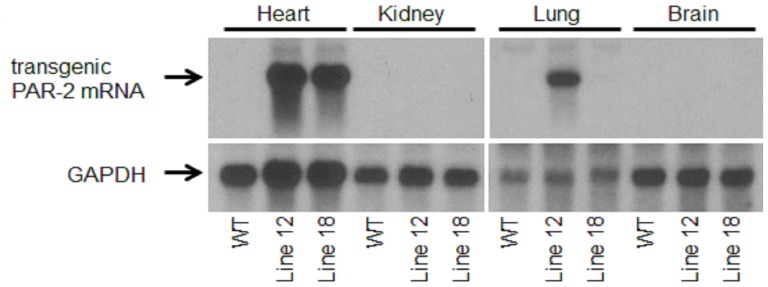
Heart failure is a major clinical problem worldwide. Previous studies have demonstrated an important role for G protein-coupled receptors, including protease-activated receptors (PARs), in the pathology of heart hypertrophy and failure. Activation of PAR-2 on cardiomyocytes has been shown to induce hypertrophic growth in vitro. PAR-2 also contributes to myocardial infarction and heart remodeling after ischemia/reperfusion injury. In this study, we found that PAR-2 induced hypertrophic growth of cultured rat neonatal cardiomyocytes in a MEK1/2 and p38 dependent manner. In addition, PAR-2 activation on mouse cardiomyocytes increased expression of the pro-fibrotic chemokine MCP-1. Furthermore, cardiomyocyte-specific overexpression of PAR-2 in mice induced heart hypertrophy, cardiac fibrosis, inflammation and heart failure. Finally, in a mouse model of myocardial infarction induced by permanent ligation of the left anterior descending coronary artery, PAR-2 deficiency attenuated heart remodeling and improved heart function independently of its contribution to the size of the initial infarct. Taken together, our data indicate that PAR-2 signaling contributes to the pathogenesis of hypertrophy and heart failure.
Conflict of interest statement
Figures


Similar articles
-
Protease-activated receptor-1 contributes to cardiac remodeling and hypertrophy.Circulation. 2007 Nov 13;116(20):2298-306. doi: 10.1161/CIRCULATIONAHA.107.692764. Epub 2007 Oct 29. Circulation. 2007. PMID: 17967980 Free PMC article.
-
Ginseng reverses established cardiomyocyte hypertrophy and postmyocardial infarction-induced hypertrophy and heart failure.Circ Heart Fail. 2012 Jul 1;5(4):504-14. doi: 10.1161/CIRCHEARTFAILURE.112.967489. Epub 2012 May 10. Circ Heart Fail. 2012. PMID: 22576957
-
Overexpression of Cardiomyocyte α1A-Adrenergic Receptors Attenuates Postinfarct Remodeling by Inducing Angiogenesis Through Heterocellular Signaling.Arterioscler Thromb Vasc Biol. 2015 Nov;35(11):2451-9. doi: 10.1161/ATVBAHA.115.305919. Epub 2015 Sep 3. Arterioscler Thromb Vasc Biol. 2015. PMID: 26338300 Free PMC article.
-
The p38 mitogen-activated protein kinase pathway--a potential target for intervention in infarction, hypertrophy, and heart failure.J Mol Cell Cardiol. 2011 Oct;51(4):485-90. doi: 10.1016/j.yjmcc.2010.10.021. Epub 2010 Nov 6. J Mol Cell Cardiol. 2011. PMID: 21062627 Free PMC article. Review.
-
Protease-activated receptors and myocardial infarction.IUBMB Life. 2011 Jun;63(6):383-9. doi: 10.1002/iub.441. Epub 2011 Mar 24. IUBMB Life. 2011. PMID: 21438116 Free PMC article. Review.
Cited by
-
Role of the protease-activated receptor-2 (PAR2) in the exacerbation of house dust mite-induced murine allergic lung disease by multi-walled carbon nanotubes.Part Fibre Toxicol. 2023 Aug 14;20(1):32. doi: 10.1186/s12989-023-00538-6. Part Fibre Toxicol. 2023. PMID: 37580758 Free PMC article.
-
Rivaroxaban attenuates cardiac hypertrophy by inhibiting protease-activated receptor-2 signaling in renin-overexpressing hypertensive mice.Hypertens Res. 2021 Oct;44(10):1261-1273. doi: 10.1038/s41440-021-00700-7. Epub 2021 Jul 20. Hypertens Res. 2021. PMID: 34285375
-
Protease-activated receptor 2 deficient mice develop less angiotensin II induced left ventricular hypertrophy but more cardiac fibrosis.PLoS One. 2024 Dec 5;19(12):e0310095. doi: 10.1371/journal.pone.0310095. eCollection 2024. PLoS One. 2024. PMID: 39637045 Free PMC article.
-
Direct oral anticoagulants and vitamin K antagonists are linked to differential profiles of cardiac function and lipid metabolism.Clin Res Cardiol. 2019 Jul;108(7):787-796. doi: 10.1007/s00392-018-1408-y. Epub 2019 Jan 2. Clin Res Cardiol. 2019. PMID: 30604046
-
Cardiac Expression of Factor X Mediates Cardiac Hypertrophy and Fibrosis in Pressure Overload.JACC Basic Transl Sci. 2020 Jan 27;5(1):69-83. doi: 10.1016/j.jacbts.2019.10.006. eCollection 2020 Jan. JACC Basic Transl Sci. 2020. Retraction in: JACC Basic Transl Sci. 2022 Sep 26;7(9):970-971. doi: 10.1016/j.jacbts.2022.08.001. PMID: 32043021 Free PMC article. Retracted.
References
-
- Lloyd-Jones D, Adams R, Carnethon M, De Simone G, Ferguson TB, et al. (2009) Heart disease and stroke statistics–2009 update: a report from the American Heart Association Statistics Committee and Stroke Statistics Subcommittee. Circulation 119: 480–486. - PubMed
-
- Hunt SA, Abraham WT, Chin MH, Feldman AM, Francis GS, et al. (2005) ACC/AHA 2005 Guideline Update for the Diagnosis and Management of Chronic Heart Failure in the Adult: a report of the American College of Cardiology/American Heart Association Task Force on Practice Guidelines (Writing Committee to Update the 2001 Guidelines for the Evaluation and Management of Heart Failure): developed in collaboration with the American College of Chest Physicians and the International Society for Heart and Lung Transplantation: endorsed by the Heart Rhythm Society. Circulation 112: e154–235. - PubMed
-
- Brown RD, Ambler SK, Mitchell MD, Long CS (2005) The cardiac fibroblast: therapeutic target in myocardial remodeling and failure. Annu Rev Pharmacol Toxicol 45: 657–687. - PubMed
-
- Chien KR, Olson EN (2002) Converging pathways and principles in heart development and disease: CV@CSH. Cell 110: 153–162. - PubMed
-
- Jessup M, Brozena S (2003) Heart failure. N Engl J Med 348: 2007–2018. - PubMed
Publication types
MeSH terms
Substances
Grants and funding
LinkOut - more resources
Full Text Sources
Other Literature Sources
Medical
Molecular Biology Databases
Miscellaneous
