. 2013 Dec 3;8(12):e80737.
doi: 10.1371/journal.pone.0080737. eCollection 2013.

PRICKLE1 interaction with SYNAPSIN I reveals a role in autism spectrum disorders

Affiliations

PRICKLE1 interaction with SYNAPSIN I reveals a role in autism spectrum disorders

Lily Paemka et al. PLoS One. .

Abstract

The frequent comorbidity of Autism Spectrum Disorders (ASDs) with epilepsy suggests a shared underlying genetic susceptibility; several genes, when mutated, can contribute to both disorders. Recently, PRICKLE1 missense mutations were found to segregate with ASD. However, the mechanism by which mutations in this gene might contribute to ASD is unknown. To elucidate the role of PRICKLE1 in ASDs, we carried out studies in Prickle1(+/-) mice and Drosophila, yeast, and neuronal cell lines. We show that mice with Prickle1 mutations exhibit ASD-like behaviors. To find proteins that interact with PRICKLE1 in the central nervous system, we performed a yeast two-hybrid screen with a human brain cDNA library and isolated a peptide with homology to SYNAPSIN I (SYN1), a protein involved in synaptogenesis, synaptic vesicle formation, and regulation of neurotransmitter release. Endogenous Prickle1 and Syn1 co-localize in neurons and physically interact via the SYN1 region mutated in ASD and epilepsy. Finally, a mutation in PRICKLE1 disrupts its ability to increase the size of dense-core vesicles in PC12 cells. Taken together, these findings suggest PRICKLE1 mutations contribute to ASD by disrupting the interaction with SYN1 and regulation of synaptic vesicles.


Conflict of interest statement

Competing Interests: The authors have declared that no competing interests exist.

Figures

Figure 1
Figure 1. Prickle1+/− mice exhibited abnormal activity consistent with ASD-like behavior.
(a) Open field chambers were used to carry out freely moving social assays to measure sociability in the Prickle1+/− mice. Eleven Prickle1+/− and sixteen wild-type C57/BL6 adult mice at 8–12 weeks old were tested. An unfamiliar, age-matched, wild-type mouse habituated the test chamber for 10 minutes. Each test mouse was then introduced and videotaped for 10 minutes. Sociability was scored by the amount of time the test mouse spent inspecting the novel mouse by sniffing, close huddling, and crawling over the other mouse. Compared to the WT, heterozygous mice exhibited deficiencies in social behavior (**p-value = 0.00084). (b) Prickle1+/− mice exhibited altered circadian rhythms. The home cage monitoring system, equipped with an activity sensor was used in this assay. Six each of WT and heterozygote Prickle1+/− littermates were tested. The activity sensor linked to a computer program detected and measured motor activity when the test mouse moved. Motions such as horizontal locomotion, rearing, climbing on the lid, grooming, and other fine movements were recorded continuously using a CCD camera and infrared illuminations for 24 hours. During the active phase, Prickle1+/− mice were significantly more active than the WT (***p-value = 0.0000002417); however, in the rest phase Prickle1+/− mice were significantly less active than their WT counterparts (**p-value = 0.00036).
Figure 2
Figure 2. PRICKLE1 Yeast two-hybrid screen isolates Unknown Short Interacting Prickle1 Peptide (USIPP).
Using Human PRICKLE1 (aa 1 to 827) N-LexA-PRICKLE1-C cloned into pB27 vector as bait, a yeast two-hybrid assay was used to screen random-primed adult and fetal cDNA libraries constructed into the pP6 vector. 53 million clones (adult brain library) and 99 million clones (fetal brain library) were screened using a mating approach and nutritional selection in media lacking tryptophan, leucine, and histidine. Applying a high Predicted Biological Score (PBS), nine final sequences were identified out of a total of 507 positive clones selected. Multiple sequence alignment of the peptides with Kalign® highlights an unknown 42-residue consensus peptide sequence (USIPP).
Figure 3
Figure 3. Coimmunoprecipitation of full-length and N-terminus PRICKLE1 with USIPP.
(a) Flag-tagged Full-length PRICKLE1 (aa 1 to 831), PRICKLE1 N-terminus (aa 1 to 313) and PRICKLE1 C-terminus (aa 314 to 831). The N-terminus includes the PET/LIM domains: a region known to mediate PRICKLE1 protein-protein interactions. (b) HEK293 cells were co-transfected with the indicated constructs (Flag-PRICKLE1+ GFP-USIPP, Flag-NPRICKLE1+ GFP-USIPP, Flag-CPRICKLE1+ GFP-USIPP or Flag-PRICKLE1+ GFP) and lysed after a 48-hour incubation period in NET-100 buffer. Lysates were immunoprecipitated with agarose-conjugated anti-GFP beads and eluted in Laemmli buffer. Immunoprecipitates were resolved by SDS-PAGE and subjected to anti-flag Western blot analysis.
Figure 4
Figure 4. USIPP antibody recognizes a brain-specific, 74 kDa protein.
(a) Anti-USIPP immunoblot shows an unknown brain-specific 74 kDa in a wild-type mouse. Lysates prepared from wild-type mouse brain, kidney or liver were resolved by SDS-PAGE and subjected to anti-USIPP Western blot analysis. (b) Confocal images show USIPP antibody immunostaining endogenous protein in dentate gyrus region of a wild-type mouse hippocampus. Sections of WT mouse hippocampi were incubated in anti-PSD-95 and rabbit pre-immune serum or immune USIPP serum followed by red AlexaFluor568 goat anti-mouse (PSD-95) and green AlexaFluor488 goat anti-rabbit (USIPP) secondary antibodies. Confocal images were captured with a Zeiss 710 microscope. The size markers correspond to 100 µm.
Figure 5
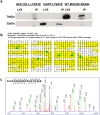
Figure 5. Protein immunoprecipitated with anti-USIPP antibodies identified as SYNAPSIN I.
(a) Wild-type mouse brain was lysed in ice-cold tissue lysis buffer. Lysates were immunoprecipitated overnight with A/G beads and anti-USIPP antibodies. The immunoprecipitate was resolved and verified with anti-USIPP Western blotting. (b) Immunoprecipitated protein was analyzed using mass spectrometry. Thirteen unique peptides that matched SYNAPSIN I protein were identified. The 13 unique proteins identified by the mass spectrometer represented 59% coverage of SYN1. (c) Representative results of the amino acid AMUs and identified mass-to-ion ratio chromatogram of a single identified peptide of SYNAPSIN I region homologous to USIPP.
Figure 6
Figure 6. USIPP antibodies recognizes human SYNAPSIN 1A.
(a) Myc-DDK- SYNAPSIN IA, Myc-REST, GFP-USIPP plasmids were transfected into HEK293 cells, incubated for 48 hours and lysed in NET-100 buffer. The lysates together with appropriate controls were resolved by SDS-PAGE and subjected to Western blot analysis with anti-USIPP or anti-Myc antibodies. (b) USIPP/SYN1 homology region, SYN1 D-DOMAIN and mutations implicated in autism and epilepsy. Multiple protein sequence alignment of human SYN1, murine SYN1 and USIPP with ClustalW shows a 31% USIPP identity with human and murine Synapsin1 from aa 431 to 483 in the D-domain; mutations shown in this domain have been implicated in autism and epilepsy. Antigen used for generating the anti-USIPP antibody is underlined in illustration. (c) USIPP antibody recognizes human Synapsin1a/USIPP homology region. USIPP/Syn1 homology region (aa 431 to 483 of SYNAPSIN I; eGFP-ΔhSyn1) was GFP-tagged and cloned into the pcDNA3.1 vector. Lysates from HEK293 cells transfected with Clones of eGFP-ΔhSyn1, (X & Y), GFP-USIPP or full-length Myc-hSyn1 were resolved with SDS-PAGE and subjected to anti-USIPP Western blot analysis.
Figure 7
Figure 7. Endogenous SYN co-localizes with PRICKLE in the mouse brain and Drosophila neuromuscular junction, and coimmunoprecipitates with PRICKLE1 in mouse brain.
(a) Co-localization of Prickle1 with Synapsin I in mouse hippocampal neurons in primary culture. Merged fluorescence image shows endogenous expression of PRICKLE1 (green) and SYNAPSIN I (red). Hippocampal neurons were fixed 10 days post-culture and immunostained with PRICKLE1 and SYNAPSIN I primary antibodies followed by AlexaFluor568 (Synapsin1) and AlexaFluor488 (Prickle1) secondary antibodies (b) EGFP-Pk and Synapsin co-localize at the neuromuscular junction (NMJ) in Drosophila third instar larvae. (I–III) 40X or (I′–III′) 100X immunohistochemistry confocal images of EGFP-Pk (I and I′) and Synapsin (II and II′) visualized at synaptic boutons of larval NMJs, along with the relevant merged images (III and III′). Note that there is a substantial co-localization of the EGFP-Pk and Synapsin signals. Scale bars = 5 µm (40X) and 5 µm (100X). Anti-GFP = rabbit anti-GFP Anti-Synapsin = mouse anti-Synapsin. (c) Lysates prepared from wild-type mouse brain was incubated with A/G agarose beads and USIPP antibodies, pre-immune serum or no serum overnight. Immunoprecipitates eluted in Laemmli buffer, resolved by SDS-PAGE and subjected to Western blot analyses with anti-PRICKLE1. (d) Lysates prepared from wild-type mouse brain was incubated with A/G agarose beads and Prickle1 antibodies, pre-immune serum or no serum overnight. Immunoprecipitates eluted in Laemmli buffer, resolved by SDS-PAGE and subjected to Western blot analyses with anti-Synapsin I.
Figure 8
Figure 8. Mutant Prickle1 exhibits altered activity in inducible stable PC12 cells.
(a) Fluorescence microscopy shows doxycycline (dox) induction of GFP in PC12 (Panel II) and differentiation in the presence of NGF after a 72-hr incubation period (Panel III). The size bars correspond to 20 nm. (b) Anti-GFP Western blot shows expression of stably transfected PC612 cells expressing GFP, GFP-PK1 or GFP-PK1R104Q under the control of dox-regulatable promoters. Lysates from dox-treated and untreated cell lines were resolved by SDS-PAGE and subjected to anti-GFP Western blot analysis. Image shows dox induction of transgenes. Anti-β actin Western Blot served as the loading control. (c) Transmission Electron Microscope (TEM) images of differentiated dox-treated and untreated PC12 cells, expressing GFP, WT, and mutant PRICKLE1, showing ultrastructure of Dense Core Vesicles (DCVs) in cytoplasm. The size bars correspond to 300 nm. Cell lines were differentiated with Nerve Growth Factor (NGF) with or without dox, for 7 days. (d) Using ImageJ, the average surface areas of DVCs in differentiated dox-treated were calculated to assess the effect of GFP, GFP-PK1 or GFP-PK1R104Q. Compared to the GFP control (43.22+/−13.78 nm2), wild-type PRICKLE1 significantly increased vesicle size (51.94+/−23.93 nm2, *p-value = 0.037) whereas mutant Prickle1 significantly decreased vesicle size (30.69+/−9.21 nm2,**p-value<0.005). The difference in activity between the wild-type and mutant Prickle1 was significant (***p-value<0.005).

References

    1. Lord C, Cook EH, Leventhal BL, Amaral DG (2000) Autism spectrum disorders. Neuron 28: 355–363. - PubMed
    1. Bailey A, Phillips W, Rutter M (1996) Autism: towards an integration of clinical, genetic, neuropsychological, and neurobiological perspectives. J Child Psychol Psychiatry 37: 89–126. - PubMed
    1. Randolph-Gips M, Srinivasan P (2012) Modeling autism: a systems biology approach. J Clin Bioinforma 2: 17. - PMC - PubMed
    1. Smalley SL (1997) Genetic influences in childhood-onset psychiatric disorders: autism and attention-deficit/hyperactivity disorder. Am J Hum Genet 60: 1276–1282. - PMC - PubMed
    1. Dravet C (2002) [The behavioral disorders in epilepsy]. Revue neurologique 158: 4S33–38. - PubMed

Publication types

MeSH terms

LinkOut - more resources