. 2014 Mar;39(5):757-70.
doi: 10.1111/ejn.12450. Epub 2013 Dec 9.

Re-evaluation of connexins associated with motoneurons in rodent spinal cord, sexually dimorphic motor nuclei and trigeminal motor nucleus

Affiliations

Re-evaluation of connexins associated with motoneurons in rodent spinal cord, sexually dimorphic motor nuclei and trigeminal motor nucleus

W Bautista et al. Eur J Neurosci. 2014 Mar.

Abstract

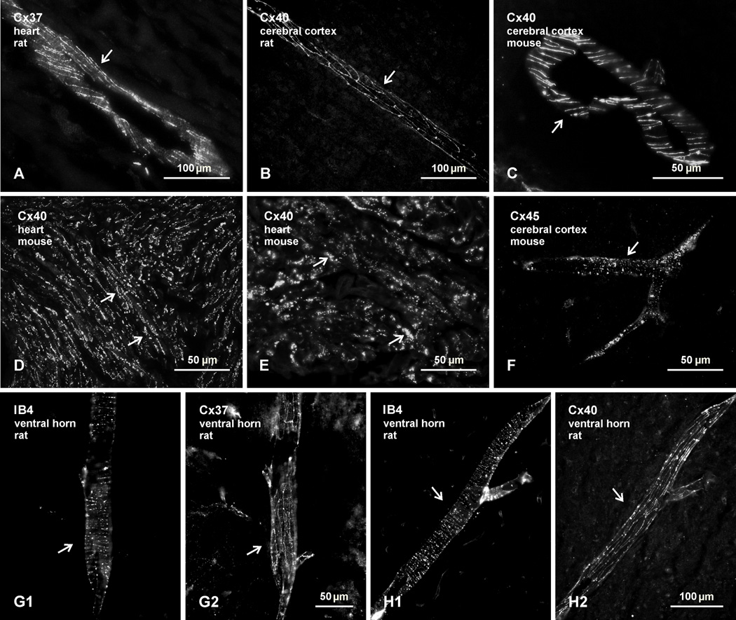
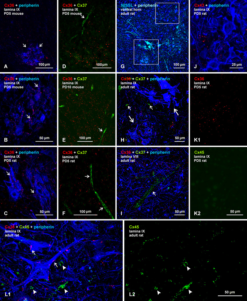
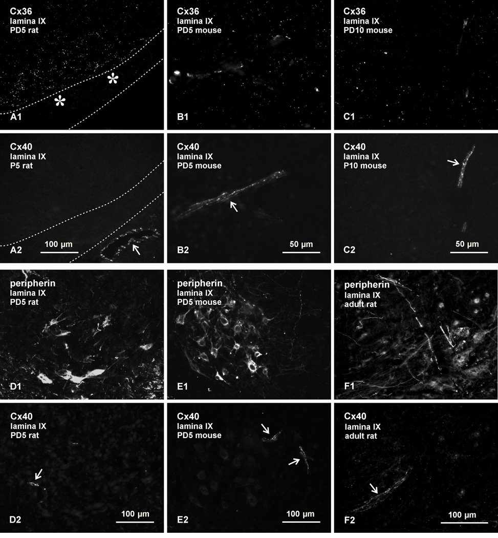
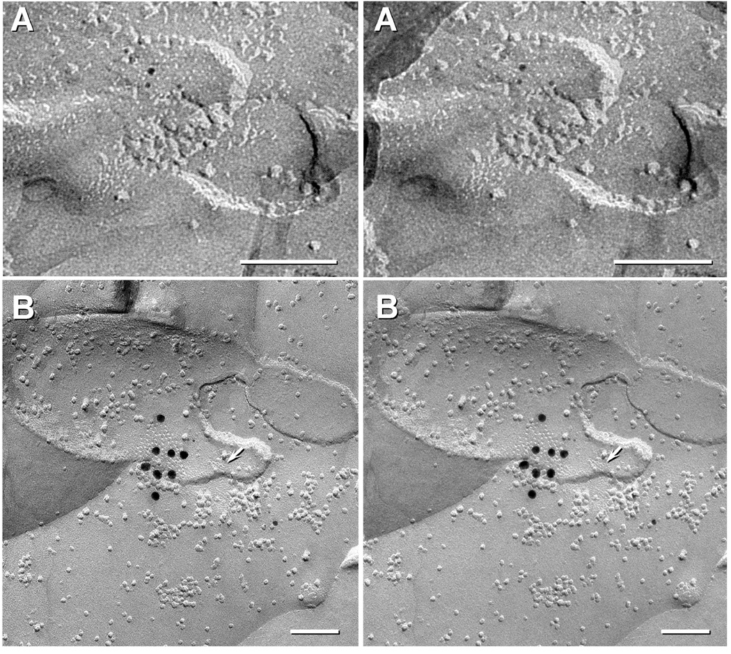
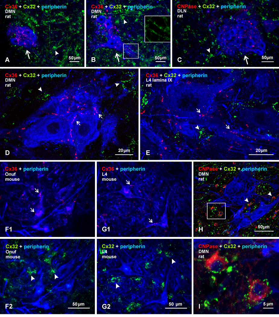
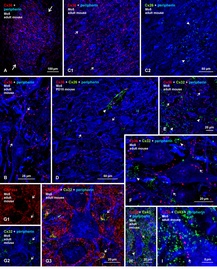
Electrical synapses formed by neuronal gap junctions composed of connexin36 (Cx36) are a common feature in mammalian brain circuitry, but less is known about their deployment in spinal cord. It has been reported based on connexin mRNA and/or protein detection that developing and/or mature motoneurons express a variety of connexins, including Cx26, Cx32, Cx36 and Cx43 in trigeminal motoneurons, Cx36, Cx37, Cx40, Cx43 and Cx45 in spinal motoneurons, and Cx32 in sexually dimorphic motoneurons. We re-examined the localization of these connexins during postnatal development and in adult rat and mouse using immunofluorescence labeling for each connexin. We found Cx26 in association only with leptomeninges in the trigeminal motor nucleus (Mo5), Cx32 only with oligodendrocytes and myelinated fibers among motoneurons in this nucleus and in the spinal cord, and Cx37, Cx40 and Cx45 only with blood vessels in the ventral horn of spinal cord, including those among motoneurons. By freeze-fracture replica immunolabeling, > 100 astrocyte gap junctions but no neuronal gap junctions were found based on immunogold labeling for Cx43, whereas 16 neuronal gap junctions at postnatal day (P)4, P7 and P18 were detected based on Cx36 labeling. Punctate labeling for Cx36 was localized to the somatic and dendritic surfaces of peripherin-positive motoneurons in the Mo5, motoneurons throughout the spinal cord, and sexually dimorphic motoneurons at lower lumbar levels. In studies of electrical synapses and electrical transmission between developing and between adult motoneurons, our results serve to focus attention on mediation of this transmission by gap junctions composed of Cx36.

Keywords: electrical coupling; gap junctions; sexually dimorphic motoneurons.


Figures

Fig. 1
Fig. 1
Confirmation of immunofluorescence labelling with anti-connexin antibodies in various tissues of adult rat and mouse. (A–C) Immunolabelling for Cx37 in rat heart (A), and Cx40 in rat (B) and mouse (C) cerebral cortex, showing detection of linear arrangements of these connexins along blood vessels (arrows), in accordance with patterns of their organization and localization at gap junctions between endothelial cells. (D,E) Labelling for Cx40 at low (D) and higher (E) magnification in atrium of mouse heart, where Cx40 is concentrated in gap junctions between lateral membranes of cardiomyocytes (D, arrows) and in gap junctions at intercalated discs (E, arrows). (F) Labelling of Cx45 along a blood vessel in mouse cerebral cortex (arrow), reflecting association of Cx45-puncta with vascular smooth muscle cells. (G,H) Images showing blood vessels in the spinal cord ventral horn labelled for the vessel marker IB4 (G1,H1, arrows) and images of the same vessels labelled for either Cx37 (G2, arrow) or Cx40 (H2, arrow), respectively, confirming vascular localization of the two connexins in spinal cord.
Fig. 2
Fig. 2
Comparison of immunofluorescence labelling for Cx36, Cx37, Cx43 and Cx45 among lumbar spinal motoneurons in lamina IX of neonatal and adult mouse and rat. In all figures, color code for secondary antibody fluorochrome and target protein is as indicated. (A–C) Images showing Cx36-puncta among peripherin-positive motoneurons (A, arrows) in mouse spinal cord at PD5, and higher magnification showing association of Cx36-puncta with motoneuron somata and dendrites in mouse (B, arrows) and rat (C, arrows) spinal cord at PD5. (D–F) Double immunofluorescence for Cx36 and Cx37 among motoneurons (peripherin labelling excluded), showing widely distributed Cx36-puncta in mouse at PD5 (D) and PD10 (E), and in rat at PD5 (F), with labelling for Cx37 restricted to blood vessels (arrows). (G) Adult rat spinal cord ventral horn labelled for peripherin and counterstained with blue fluorescence Nissl. (H,I) Magnifications of boxed areas in G (H, lower box; I, upper box) triple labelled for Cx36, Cx37 and peripherin, showing Cx36-puncta among two peripherin-positive motoneuronal groups (H, large arrows), absence of labelling for Cx37 among these groups, and labelling of Cx37 restricted to small blood vessels (H, small arrows) and a single large vessel in lamina VIII (I, arrow). (J) Double labelling of Cx43 with peripherin in lamina IX of rat at PD5, showing very little association of sparsely-distributed Cx43-puncta with the surface of peripherin-positive motoneurons. (K) Double labelling of Cx36 with Cx45 in lamina IX of rat at PD5, showing Cx36-puncta among motoneurons (K1) and only a low level of background fluorescence in the same field labelled for Cx45 (K2). (L) Triple labelling for Cx36, Cx45 and peripherin in lamina IX of adult rat, showing Cx36-puncta on the surface of peripherin-positive motoneurons (L1, arrows), minimal Cx45 localization to these neurons, and association of Cx45 with what appear to be blood vessels (L1, arrowheads), also shown in the same field with labelling of Cx45 alone (L2).
Fig. 3
Fig. 3
Immunofluorescence labelling for Cx36, Cx40 and peripherin in lamina IX of lumbar spinal cord in rat and mouse. Double labelling is shown in the same fields in pairs of images: A1,A2; B1,B2, etc, through to F1,F2. (A–C) Fields displaying punctate labelling for Cx36 (A1, B1 and C1) in lamina IX show labelling for Cx40 only along blood vessels in rat at PD5 (A2, arrow), and in mouse at PD5 (B2, arrow) and at PD10 (C2, arrow). The image in (A) shows a portion of ventral white matter (delineated by dotted lines) to allow inclusion of an underlying blood vessel as a positive control for labelling of Cx40. (D,E) Labelling of peripherin (D1,E1) and Cx40 (D2,E2) in lamina IX, showing absence of labelling for Cx40 specifically within or among peripherin-positive motoneurons, and association of Cx40 with blood vessels (arrows) in rat (D) and mouse (E) at PD5. (F) Labelling of peripherin (F1) and Cx40 (F2) in lamina IX of adult rat, showing absence of labelling for Cx40 in association with peripherin-positive motoneurons, and association of Cx40 with a blood vessel (F2, arrow).
Fig. 4
Fig. 4
Stereoscopic FRIL images of neuronal vs. astrocyte gap junctions in developing rat spinal cord. (A) Small neuronal gap junction in P7 spinal cord labelled for Cx36 by three 5-nm gold beads but not labelled for Cx43 (10-nm and 20-nm gold; none present). (B) A medium-size astrocyte gap junction from P4 spinal cord labelled for Cx43 by eight 18 nm gold beads. This replica was also labelled for AQP4 (6 and 12nm gold beads). Arrow points to an E-face imprint of a square array, clearly marking this as an A/A gap junction. Calibration bars are 0.1 micron.
Fig. 5
Fig. 5
Immunofluorescence labelling for Cx36, Cx32, and peripherin in sexually dimorphic and non-dimorphic lumbar motor nuclei in adult male rat and mouse spinal cord. (A–C) Images from rat showing rostral (A) and caudal (B) regions of the dimorphic dorsomedial nucleus (DMN), and the dorsolateral nucleus (DLN), shown in (C). Cx36-puncta are seen among clusters of peripherin-positive motoneurons (arrows), and labelling for Cx32 is seen largely in surrounding regions (arrowheads). Boxed area (B, lower box; magnified in inset of upper box, without labelling for peripherin) shows Cx32 localized to myelinated fibers passing between motoneuron dendrites. (D,E) Higher magnifications showing Cx36-puncta localized to the surface of peripherin-positive motoneurons in the dorsomedial dimorphic nucleus (D, arrows) and in a non-dimorphic motor nucleus at L4 (E, arrows), with little Cx32 (arrowheads) association at motoneurons. Cx36-puncta follow apposition of two neuronal somata (D, left arrow). (F) Images of sexually dimorphic Onuf’s nucleus in mouse, showing Cx36-puncta associated with peripherin-positive Onuf motoneurons (F1, arrows) and, in the same field, labelling for Cx32 localized to small cells among these motoneurons (F2, arrowheads). (G) Image of non-dimorphic motor nucleus from L4 of mouse, showing a similar pattern of Cx36-puncta associated with motoneurons (G1, arrows), and labelling for Cx32 at small cells (G2, arrowheads). (H,I) Triple labelling for Cx32, CNPase and peripherin in the dorsomedial dimorphic nucleus at low magnification (H), with boxed area in H magnified in (I), showing Cx32 localized to CNPase-positive oligodendrocytes (arrowheads) and not to peripherin-positive motoneurons.
Fig. 6
Fig. 6
Immunofluorescence labelling of Cx36, Cx26, Cx32, Cx43, peripherin and CNPase in the trigeminal motor nucleus (Mo5) of adult and PD15 mouse. (A) Low magnification showing a high density of labelling for Cx36 among peripherin-positive motoneurons in adult mouse Mo5 (large arrows) and sparse labelling in surrounding regions. (B) Magnification showing punctate appearance of labelling for Cx36, association of Cx36-puncta with motoneuron somata and dendrites (arrows), and absence of intracellular labelling for Cx36. (C) Triple immunofluorescence showing overlay of labelling for Cx36 with peripherin and, in the same field, overlay of labelling for Cx26 and peripherin. Cx36-puncta are seen throughout the field, including those associated with medially located motoneurons (C1, arrows), while Cx26-puncta are seen restricted to the lateral edge of the Mo5, in a region largely devoid of peripherin-positive motoneuron somata, but containing bundles of peripherin-positive myelinated fibers in the motor root of the trigeminal nerve (C2, arrowheads). (D) Triple labelling in Mo5 at PD15, showing Cx36-puncta decorating motoneuron somata and dendrites (arrows), absence Cx26 labelling in or around these somata, and Cx26-puncta associated with leptomeninges along a blood vessel (arrowhead). (E,F) Low (E) and higher (F) magnification showing labelling of Cx36 and Cx32 among peripherin-positive Mo5 motoneurons. Cx36-puncta are seen on the surface (confirmed by confocal through focus analysis) of peripherin-positive motoneurons (arrows), while Cx32-puncta are largely in regions devoid of peripherin labelling (arrowheads). (G) Triple labelling, showing CNPase localized to oligodendrocyte somata (G1, arrows) and myelinated fibers, and localization of Cx32-puncta to CNPase-positive oligodendrocyte somata (G2, arrows), as shown in overlay (G3, arrows). (H,I) Low (H) and higher (I) magnification showing overlay of triple labelling for Cx36 and Cx43 among peripherin-positive Mo5 motoneurons. Dense Cx43-puncta are seen in the vicinity of motoneurons (arrowhead), and display a random distribution compared with lining of Cx36-puncta on the surface of motoneurons (H,I, arrows). Motoneurons lack intracellular labelling for Cx43 (confirmed by confocal through focus analysis).

References

    1. Arasaki K, Kudo N, Nakanishi T. Firing of spinal motoneurons due to electrical interactions in the rat: an in vitro study. Exp. Brain Res. 1984;54:437–445. - PubMed
    1. Bautista W, Nagy JI, Dai Y, McCrea DA. Requirement of neuronal connexin36 in pathways mediating presynaptic inhibition of primary afferents in functionally mature mouse spinal cord. J Physiol. 2012;590:3821–3839. - PMC - PubMed
    1. Bautista W, Nagy JI. Connexin36 in gap junctions forming electrical synapses between motoneurons in sexually dimorphic motor nuclei in spinal cord of rat and mouse. Neuroscience. 2013a In Press. - PMC - PubMed
    1. Bautista W, McCrea DA, Nagy JI. Connexin36 identified at primary afferent terminals forming morphologically mixed chemical/electrical synapses in adult rodent spinal cord. 2013b Submitted. - PMC - PubMed
    1. Bennett MVL. Gap junctions as electrical synapses. J. Neurocytol. 1997;26:349–366. - PubMed

Publication types