. 2014 Jan 5:448:293-302.
doi: 10.1016/j.virol.2013.10.018. Epub 2013 Nov 8.

KSHV LANA and EBV LMP1 induce the expression of UCH-L1 following viral transformation

Affiliations

KSHV LANA and EBV LMP1 induce the expression of UCH-L1 following viral transformation

Gretchen L Bentz et al. Virology. .

Abstract

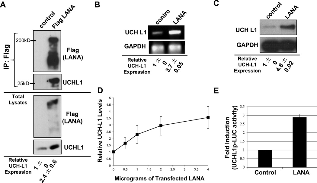
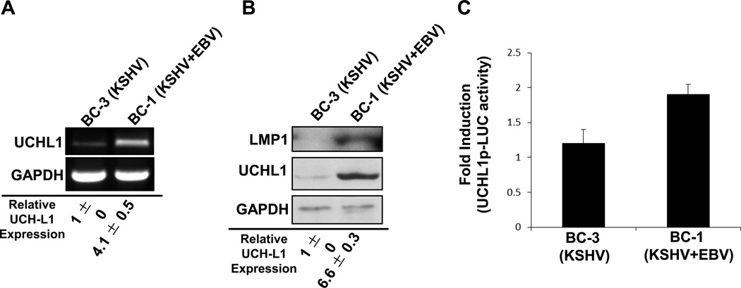
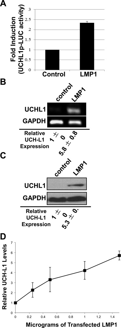
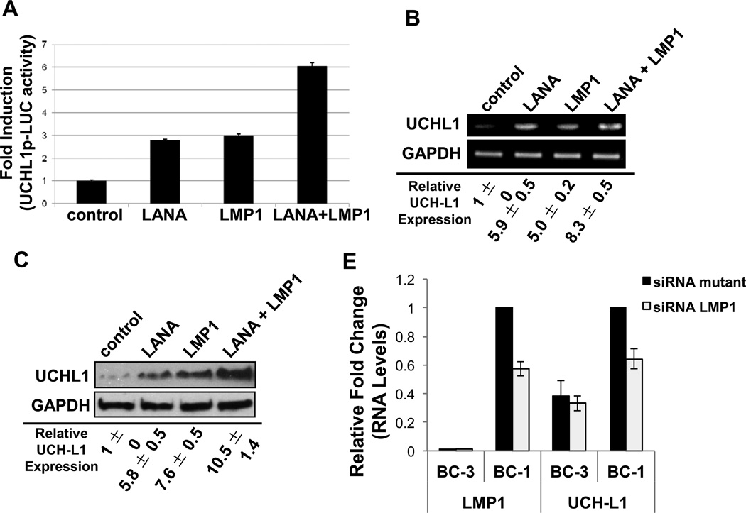
Ubiquitin C-terminal Hydrolase L1 (UCH-L1) has oncogenic properties and is highly expressed during malignancies. We recently documented that Epstein-Barr virus (EBV) infection induces uch-l1 expression. Here we show that Kaposi's Sarcoma-associated herpesvirus (KSHV) infection induced UCH-L1 expression, via cooperation of KSHV Latency-Associated Nuclear Antigen (LANA) and RBP-Jκ and activation of the uch-l1 promoter. UCH-L1 expression was also increased in Primary Effusion Lymphoma (PEL) cells co-infected with KSHV and EBV compared with PEL cells infected only with KSHV, suggesting EBV augments the effect of LANA on uch-l1. EBV latent membrane protein 1 (LMP1) is one of the few EBV products expressed in PEL cells. Results showed that LMP1 was sufficient to induce uch-l1 expression, and co-expression of LMP1 and LANA had an additive effect on uch-l1 expression. These results indicate that viral latency products of both human γ-herpesviruses contribute to uch-l1 expression, which may contribute to the progression of lymphoid malignancies.

Keywords: EBV; KSHV; LANA; LMP1; UCH-L1.


Figures

Figure 1
Figure 1. UCH-L1 expression is induced in endothelial cells after infection with KSHV
(A) Equivalent amounts of HUVEC and KSHV-HUVEC cell lysates were subjected to 2D gel electrophoresis analysis following which the gels were stained with Coomassie blue. Two of the differentially expressed spots, identified by mass spectrometry, were UCH-L1 and GFP (arrows). (B) RNA from HUVEC and KSHV-HUVEC was isolated and subjected to RT-PCR using uch-l1 and β-actin primers. (C) Equivalent amounts of KSHVHUVEC and HUVEC cell lysates were subjected to SDS-PAGE and immunoblotted with UCH-L1 or actin antibodies. Relative expression was determined by densitometry. All results are shown as the mean ± standard deviation for experiments performed in triplicate.
Figure 2
Figure 2. KSHV LANA is associated with endogenous UCH-L1 and induces expression of UCH-L1
(A) Cos7 cells were transfected with control or LANA-Flag expression vectors and harvested 48h post-transfection for immunoprecipitation analysis. LANA-Flag was immunoprecipitated with anti-Flag-agarose beads. IPs and cell lysates were resolved on 10–12% SDS-PAGE and probed with UCH-L1 and Flag antibodies. (B–C) Total RNA and protein were extracted from cells co-transfected with LANA or control expression constructs. (B) RT-PCR analysis was performed using primers specific for uchl1 and gapdh. (C) Western blot analyses for UCH-L1 protein levels in lysates from cells transfected with or without LANA were performed with UCH-L1 antibodies. GAPDH was used as loading control. (D) Cells were transfected with different amounts of Flag-LANA or vector-control expressing plasmids and Western blot analyses to detect UCH-L1 was performed. Relative expression was determined by densitometry, and results are shown as the mean fold change ± standard deviation for experiments performed in triplicate. (E) NIH3T3 cells were co-transfected with control or LANA-Flag expression vectors (350 ng/well) along with UCH-L1p-LUC wildtype reporter plasmid (500 ng/well) and β-gal expression constructs (250 ng/well). Luciferase assays were performed 48 h post-transfection. The data are shown as the mean ± standard deviation for three independent experiments in triplicate and normalized to β-gal activity.
Figure 3
Figure 3. Endogenous UCH-L1 expression is greater in primary effusion lymphoma cells co-infected with EBV
(A–B) Total RNA and protein were extracted from BC-3 and BC-1 cell lines. (A) RT-PCR was performed with uch-l1- and gapdh-specific primers. (B) Western blot analyses were performed with UCH-L1 and GAPDH antibodies. Relative expression was determined by densitometry. Results are shown as the mean ± standard deviation for experiments performed in triplicate. (C) Endogenous uch-l1 promoter activity was determined in BC-3 (positive for KSHV only) and BC-1 (co-infected with KSHV and EBV) PEL cells. Cells (2 × 10⁵) were nucleofected with UCH-L1p-LUC reporter and β-gal constructs. Luciferase assays were performed 48 h post-transfection. The experiments were done in triplicate and normalized to β-gal activity.
Figure 4
Figure 4. EBV LMP1 induces expression of UCH-L1
(A) NIH3T3 cells were co-transfected with control or LMP1-Flag expression vectors (250 ng/well), along with UCH-L1p-LUC wild type reporter plasmid (500 ng/well) and β-gal expression constructs (250 ng/well). Luciferase assays were performed 48 h post-transfection. The data represent three independent experiments performed in triplicate and normalized to β-gal activity. (B–C) Total RNA and protein were extracted from cells co-transfected with LMP1 or control. (B) RT-PCR was performed with specific primers for uch-l1 (gapdh was used as a control). (C) Western blot analyses for UCH-L1 protein levels in lysates from cells transfected with or without LMP1 were performed with UCH-L1 antibodies. GAPDH was the loading control. (D) Cells were transfected with different amounts of Flag-LANA or vector-control expressing plasmids and Western blot analyses to detect UCH-L1 was performed. Relative expression was determined by densitometry. Results are shown as the mean fold change ± standard deviation for experiments performed in triplicate.
Figure 5
Figure 5. KSHV LANA and EBV LMP1 have additive effects on the expression of UCH-L1
(A) NIH3T3 cells were co-transfected with control, Flag-LANA, and/or pcDNA3-LMP1 expression vectors (250 ng/well), along with UCH-L1p-LUC wild type reporter plasmid (500 ng/well) and β-gal expression constructs (250 ng/well). Luciferase assays were performed 48 h post-transfection. The data represent three independent experiments performed in triplicate and normalized to β-gal activity. (B–C) Total RNA and protein were extracted from cells co-transfected with the control, Flag-LANA, and/or pcDNA3-LMP1 expression constructs. (B) RT-PCR was performed with specific primers for uch-l1 (gapdh was used as a control). (C) Western blot analyses for UCH-L1 protein levels in lysates were performed. GAPDH was the loading control. (D) BC-1 and BC-3 cells were transfected with siRNA LMP1. Transfection with a siRNA LMP1 mutant, where two nucleotides were mutated, served as a control. RNA was harvested and RT-PCR was performed with specific primers for uch-l1 and LMP1 (gapdh was used as a control). The fold change in relative uch-l1 and LMP1 RNA levels (relative to gapdh) was determined. Results are shown as the mean ± standard deviation for experiments performed in triplicate.
Figure 6
Figure 6. Activation of the uch-l1 promoter by KSHV-LANA and EBV occurs via RBP-Jκ
(A) NIH3T3 cells were co-transfected with control or RBP-Jκ (100 ng/well) or LANA (350 ng/well) or RBP-Jκ and LANA together, along with UCHL1p-LUC wild type plasmid (500 ng/well) and β-gal expression constructs (250 ng/well). The control DNA was used as filler DNA to maintain the total amount of DNA constant. Luciferase assays were performed 48 h post-transfection. The data represent three independent experiments prepared in triplicate and normalized to β-gal activity. (B) ChIP/PCR analyses were performed to determine binding of RBP-Jκ factor to the putative partial binding sites on the UCH-L1 promoter with the use of specific RBP-Jκ antibody in KR4 LCLs. Normal IgG was used as negative control. PCR reactions were performed with primers targeting the partial RBP-Jκ-binding sites (see Materials and Methods), and amplified DNA products were resolved in 2% agarose gels.
Figure 7
Figure 7. Proposed Model of the Induction of uch-l1 following transformation of B cells by KSHV and/or EBV
Naïve B cells are infected and transformed by KSHV and/or EBV. EBV-mediated transformation results in EBNA2 and LMP1 expression, resulting in the activation of the uch-l1 promoter via PU.1 and RBP-Jκ, respectively, and elevated UCH-L1 expression. KSHV-mediated transformation results in LANA expression, resulting in the activation of the uch-l1 promoter via RBP-Jκ and elevated UCH-L1 expression. Transformation mediated by a dual KSHV/EBV infection results in expression of KSHV LANA and EBV LMP1. Both viral proteins independently activate the uch-l1 promoter, at least in part through RBP-Jκ, resulting in enhanced activation of the uch-l1 promoter and an additive elevation of UCH-L1 expression. Known tumorigenic phenotypes associated with elevated UCH-L1 expression include increased cell proliferation, migration, invasion, and adhesion as well as changes in cell morphology and inhibition of apoptosis.

References

    1. Basseres E, Coppotelli G, Pfirrmann T, Andersen JB, Masucci M, Frisan T. The ubiquitin C-terminal hydrolase UCH-L1 promotes bacterial invasion by altering the dynamics of the actin cytoskeleton. Cellular microbiology. 2010;12:1622–1633. - PubMed
    1. Bentz GL, Shackelford J, Pagano JS. Epstein-Barr virus latent membrane protein 1 regulates the function of interferon regulatory factor 7 by inducing its sumoylation. J Virol. 86:12251–12261. - PMC - PubMed
    1. Bentz GL, Whitehurst CB, Pagano JS. Epstein-Barr virus latent membrane protein 1 (LMP1) C-terminal-activating region 3 contributes to LMP1-mediated cellular migration via its interaction with Ubc9. J Virol. 2011;85:10144–10153. - PMC - PubMed
    1. Betarbet R, Sherer TB, Greenamyre JT. Ubiquitin-proteasome system and Parkinson's diseases. Experimental neurology. 2005;191(Suppl 1):S17–S27. - PubMed
    1. Bheda A, Gullapalli A, Caplow M, Pagano JS, Shackelford J. Ubiquitin editing enzyme UCH L1 and microtubule dynamics: implication in mitosis. Cell cycle (Georgetown, Tex. 2010;9:980–994. - PMC - PubMed

Publication types

MeSH terms

Substances