Review
. 2013 Dec 4;80(5):1129-44.
doi: 10.1016/j.neuron.2013.10.030.

A role for correlated spontaneous activity in the assembly of neural circuits

Affiliations
Review

A role for correlated spontaneous activity in the assembly of neural circuits

Lowry A Kirkby et al. Neuron. .

Erratum in

  • Neuron. 2014 Jan 8;81(1):218

Abstract

Before the onset of sensory transduction, developing neural circuits spontaneously generate correlated activity in distinct spatial and temporal patterns. During this period of patterned activity, sensory maps develop and initial coarse connections are refined, which are critical steps in the establishment of adult neural circuits. Over the last decade, there has been substantial evidence that altering the pattern of spontaneous activity disrupts refinement, but the mechanistic understanding of this process remains incomplete. In this review, we discuss recent experimental and theoretical progress toward the process of activity-dependent refinement, focusing on circuits in the visual, auditory, and motor systems. Although many outstanding questions remain, the combination of several novel approaches has brought us closer to a comprehensive understanding of how complex neural circuits are established by patterned spontaneous activity during development.


Figures

Figure 1
Figure 1. Retinal, cochlear and spinal cord projections to their primary targets
(A) Schematic representation of retinal ganglion cell (RGC) projections to their primary targets: the dorsal lateral geniculate nucleus (dLGN) of the thalamus and the superior colliculus (SC). Red areas in the dLGN and SC correspond to projections from the left (red) eye; blue areas correspond to projections from the right (blue) eye. Shading represents retinotopy. V: ventral, D: dorsal, T: temporal, N: nasal, A: anterior, P: posterior, M: medial, L: lateral. (B) Schematic representation of cochlear projections to primary brainstem targets: the cochlear nucleus (CN), the medial superior olive (MSO), the lateral superior olive (LSO) and the medial nucleus of the trapezoid body (MNTB). Red areas correspond to projections from the left (red) cochlea; blue areas correspond to projections from the right (blue) cochlea. Shading represents tonotopy. Both the LSO and MSO receive overlapping tonotopic maps originating from either cochlea. HF: high frequency, LF: low frequency. (Adapted from Kandler, 2009). (C) Schematic representation of some spinal cord cell types and their connections. MN: motoneuron, DRG: dorsal root ganglia, RC: Renshaw cell; 1a: 1a inhibitory interneuron, dC: commissural interneurons; D: dorsal, V: ventral.
Figure 2
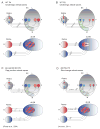
Figure 2. Retinotopic map formation and eye-specific segregation under normal and disrupted spontaneous retinal activity patterns
Schematic representations of retinotopic map formation in the SC and eye-specific segregation in the dLGN during normal development (A—B) and as a result of experiments that alter the spatial and temporal pattern of afferent RGC activity (C—H), as described in the text. Shading corresponds to retinotopy. Striped regions correspond to unsegregated inputs from left and right eyes. SC: superior colliculus, dLGN: dorsal lateral geniculate nucleus, V: ventral, D: dorsal, T: temporal, N: nasal, A: anterior, P: posterior, M: medial, L: lateral, R: right eye, L: left eye, ChR2: channelrhodopsin-2.
Figure 3
Figure 3. Motoneuron pathfinding in the developing spinal cord under normal and disrupted activity patterns
Schematic representation of motoneuron pathfinding in the developing spinal cord during normal development (A), in the presence of the GABA-A antagonist picrotoxin (B), and in the combination of picrotoxin and ChR2 stimulation (C). V: ventral, D: dorsal, MN: motoneuron, RC: Renshaw cell, 1a: 1a inhibitory interneuron; ChR2: channelrhodopsin-2.
Figure 4
Figure 4. Activity-dependent competition during eye-specific segregation in the dLGN under normal and disrupted glutamate release and targeting
Schematic representations of activity-dependent competition during eye-specific segregation in the dLGN for normal development (A), glutamate release-deficient ipsilateral projecting axons (B) and for a subset of ipsilateral axons that were genetically misrouted to project contralaterally (C), as described in the text. Left panels show afferent activity patterns; middle panels show experimental observation; right panels show prediction according to a Hebbian model of competition.
Figure 5
Figure 5. Components used in theoretical models for retinotipic refinement
Schematic representation of components used in theoretical models for retinotopic refinement: chemoaffinity gradients (A), Hebbian plasticity (B) and competition for resources (C). First, chemoaffinity gradients in the form of ephrin/Eph gradients guide RGC axons to their approximate retinotopic location and to form selective arborization. Second, a Hebbian plasticity component strengthens the synapses of cells that are driven to fire together by retinal waves. Third, competition for limited resources in the target areas function to constrain the termination zone of RGC axons. V: ventral, D: dorsal, T: temporal, N: nasal, A: anterior, P: posterior, M: medial, L: lateral. (Adapted from Grimbert and Cang, 2012).

References

    1. Ackman JB, Burbridge TJ, Crair MC. Retinal waves coordinate patterned activity throughout the developing visual system. Nature. 2012;490:219–225. - PMC - PubMed
    1. Bansal A, Singer JH, Hwang BJ, Xu W, Beaudet A, Feller MB. Mice lacking specific nicotinic acetylcholine receptor subunits exhibit dramatically altered spontaneous activity patterns and reveal a limited role for retinal waves in forming ON and OFF circuits in the inner retina. J Neurosci. 2000;20:7672–7681. - PMC - PubMed
    1. Blankenship AG, Feller MB. Mechanisms underlying spontaneous patterned activity in developing neural circuits. Nat Rev Neurosci. 2010;11:18–29. - PMC - PubMed
    1. Blumberg MS, Marques HG, Iida F. Twitching in sensorimotor development from sleeping rats to robots. Curr Biol. 2013;23:R532–R537. - PMC - PubMed
    1. Boulanger LM. Immune proteins in brain development and synaptic plasticity. Neuron. 2009;64:93–109. - PubMed