. 2013 Dec 4;80(5):1145-58.
doi: 10.1016/j.neuron.2013.09.022.

A modular gain-of-function approach to generate cortical interneuron subtypes from ES cells

Affiliations

A modular gain-of-function approach to generate cortical interneuron subtypes from ES cells

Edmund Au et al. Neuron. .

Abstract

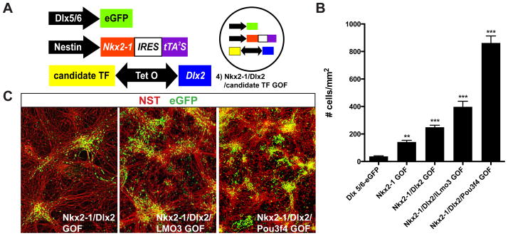
Whereas past work indicates that cortical interneurons (cINs) can be generically produced from stem cells, generating large numbers of specific subtypes of this population has remained elusive. This reflects an information gap in our understanding of the transcriptional programs required for different interneuron subtypes. Here, we have utilized the directed differentiation of stem cells into specific subpopulations of cortical interneurons as a means to identify some of these missing factors. To establish this approach, we utilized two factors known to be required for the generation of cINs, Nkx2-1 and Dlx2. As predicted, their regulated transient expression greatly improved the differentiation efficiency and specificity over baseline. We extended upon this "cIN-primed" model in order to establish a modular system whereby a third transcription factor could be systematically introduced. Using this approach, we identified Lmo3 and Pou3f4 as genes that can augment the differentiation and/or subtype specificity of cINs in vitro.


Figures

Figure 1
Figure 1. Exogenously specified ES cells become ventral telencephalic neural precursors capable of long-range tangential migration and differentiation into cortical interneurons
Embryoid bodies grown as adherent cultures express FoxG1 (A), Nkx2-1 (B), and Olig2 (C) after 9 days of differentiation. Upon dissociation, a large proportion of neurons are GABA-positive (D). (E) Schematic of transplantation strategy: A pan-eGFP expressing line was differentiated for 11 days and then dissociated and transplanted into MGE of wildtype e13.5 Swiss Webster embryos. 3 days following transplantation (e16.5), eGFP+ unipolar migratory cells were observed migrating away from the transplant site (F) and into the cortex (F′ and F″). Other transplant recipients were analyzed at P7 (G) and were GABA-positive, and others analyzed at P19 and co-expressed eGFP and cortical interneuron subtype markers, parvalbumin (PV) (H) and reelin (I). See also Figure S1.
Figure 2
Figure 2. Directed transcriptional specification of cortical interneurons with Nkx2-1 and Dlx2
(A) Schematic representation of transgenes introduced into a wildtype ES cell line. A parental line was produced by introducing 1) eGFP under the control of the Dlx5/6 intergenic region and a minimal promoter. To the parental line, two additional transgenes were sequentially introduced: 2) Nestin intron II with minimal promoter driving Nkx2-1-IRES-tTA2S and 3) Tetracycline response element driving Dlx2. Therefore three separate stable ES lines were established containing 1, 2 or all 3 transgenes: 1) Dlx5/6-eGFP, 2) Nkx2-1 GOF and 3) Nkx2-1/Dlx2 GOF, respectively. (B) All three lines were differentiated for 11 days as embryoid bodies and there was no observable difference in differentiation efficiency to telencephalic fate as measured by FoxG1. (C) Nkx2-1 was strongly and broadly expressed in the large majority of nestin-expressing cells of Nkx2-1 GOF and Nkx2-1/Dlx2 GOF, while in Dlx5/6-eGFP, its expression was limited to small clusters. (D) Timecourse of Dlx2 expression in differentiating embryoid bodies by western blot. Dlx5/6-eGFP and Nkx2-1GOF both begin expressing Dlx2 at day 7 and have similar expression levels. Nkx2-1/Dlx2 GOF expresses Dlx2 in a similar timecourse at elevated levels. Beta actin loading controls below each Dlx2 blot. (E) Quantification of Nkx2-1 and Dlx2 levels in embryoid bodies normalized to b-actin loading control during in vitro differentiation. Red asterisk denotes p<0.05 for Nkx2-1 GOF over Dlx5/6-eGFP. Blue asterisk denotes p<0.05 for Nkx2-1/Dlx2 GOF over Dlx5/6-eGFP. Data represented as mean ± SEM. See also Figure S2 and S4.
Figure 3
Figure 3. Transcriptional specification with Nkx2-1 and Dlx2 increases the directed production of cortical interneurons
(A) Representative images of neuronal monolayers generated from Dlx5/6-eGFP, Nkx2-1 GOF, and Nkx2-1/Dlx2 GOF lines; Neuron specific tubulin (red), eGFP (green). (B and C) Quantification of Dlx5/6-eGFP+ neurons by cell counts of 10x fields, normalized to mm2 (B) or by FACS analysis (C) demonstrate a step-wise improvement in differentiation efficiency with Nkx2-1 GOF alone and Nkx2-1/Dlx2 GOF together. Dlx5/6-eGFP (IHC n=6; FACS n=4); Nkx2-1 GOF (IHC n=5; FACS n=4); Nkx2-1/Dlx2 GOF (IHC n=5; FACS n=4) (* p<0.05; ** p<0.01; *** p<0.001) Data represented as mean ± SEM. See also Figures S2–4.
Figure 4
Figure 4. Transplantation of 3 differentiated transgenic ES lines demonstrates the effects of Nkx2-1 and Dlx2 on cortical interneuronal differentiation
Postnatal day 21 analysis of Dlx5/6-eGFP, Nkx2-1 GOF and Nkx2-1/Dlx2 GOF were transplanted separately into e13.5 MGE and host animals. (A–D) Examples of eGFP+ ES cell-derived cortical interneurons expressing parvalbumin (PV) (A and B), somatostatin (B and C), reelin-only (C), and vasointestinal peptide (D). (E and F) Examples of groups of ES-derived cINs exhibiting complex morphologies consistent with bona fide cINs. (G) Numbers of cINs present in the cortex quantified across the transgenic lines shows a step-wise increase in migration efficiency with Nkx2-1 alone and Nkx-1/Dlx2 together. (H) Quantification of cIN subtype markers expressed by transplanted cells demonstrates that Nkx2-1 strongly skews identity towards MGE-derived cINs, with no additional effects observed with the further addition of Dlx2. MGE vs. CGE cIN subtypes summarized as pie charts in (I). Dlx5/6-eGFP (n=8; 388 cells); Nkx2-1 GOF (n=7; 912 cells); Nkx2-1/Dlx2 GOF (n=6, 1281 cells) (* p<0.05; ** p<0.01; *** p<0.001) Data represented as mean ± SEM. See also Figures S5, S6 and S11.
Figure 5
Figure 5. Proper maturation of transplanted ES cells in the neocortex
A) Intrinsic electrophysiological properties of ES cell showing characteristics of a typical CGE-derived sigmoid intrinsic bursting cell (sIB). A1) A burst of action potentials at threshold (black) and a pulse at just sub-threshold depolarization (red). A2) A series of hyperpolarizing voltage steps and a larger depolarizing one which leads to a higher frequency adapting discharge. B) Electrophysiological properties of a transcriptionally-specified transplanted ES cell showing characteristics of a typical MGE-derived non-fast spiking cell (NFS). B1) Threshold action potential discharge (black) and just sub-threshold depolarization (red). B2) A series of hyperpolarizing voltage steps and a larger depolarizing one which leads to a higher frequency adapting discharge. B3) The recorded cell was filled with biocytin (green) and was immunoreactive for somatostatin (red) and weakly for reelin (blue). C) Electrophysiological properties of a transcriptionally-specified transplanted ES cell showing characteristics of a typical MGE-derived delayed fast spiking basket cell (dFS). C1) Threshold action potential discharge (black) and just sub-threshold depolarization (red). C2) A series of hyperpolarizing voltage steps and a larger depolarizing one which leads to a higher frequency non-adapting discharge. C3) Incoming excitatory postsynaptic currents (EPSCs) recorded from the same cell at -65mV. D) A morphological reconstruction of a transcriptionally-specified and recorded FS cell. The soma and dendrites are depicted in blue, whereas the axon in red. The axo-dendritic profile of the neuron displays characteristic features of FS cells, having an axon that bifurcates many times in the vicinity of the cell, bearing terminals that putatively contact surrounding other cell somata. See also Figure S7.
Figure 6
Figure 6. Using a transcriptional strategy to test the influence of candidate transcription factors revealed that LMO3 and Pou3f4 affect the subtype identity and induction of cortical interneurons
Of the twelve candidate transcription factors tested, LMO3 and Pou3f4 most robustly improved Dlx5/6-eGFP+ neuronal differentiation above that achieved compared to the Nkx2-1/Dlx2 GOF baseline. Candidate factors were introduced by utilizing the bidirectional nature of the tetracycline response element (A), which allowed both Dlx2 and an additional factor to be simultaneously expressed under the control of tTA2S. Dlx5/6-eGFP+ neurons were quantified by cell counts per 10x field, normalized to mm2 (B). Representative field of Nkx2-1/Dlx2 GOF, Nkx2-1/Dlx2/LMO3 GOFand Nkx2-1/Dlx2/Pou3f4 GOF (C); neuron specific tubulin (NST, red) and eGFP (green). Nkx2-1/Dlx2/LMO3 GOF (IHC n=4); Nkx2-1/Dlx2/Pou3f4 GOF (IHC n=5) (* p<0.05; ** p<0.01; *** p<0.001) Data represented as mean ± SEM. See also Figures S8–10.
Figure 7
Figure 7. Analysis of the LMO3 Null Mouse Confirms the Prediction that LMO3 Plays a Role in Generating PV+ Cortical Interneurons
Nkx2-1/Dlx2/LMO3 GOF line was differentiated and transplanted in utero into e13.5 MGE. Transplants were analyzed at P21 for numbers of cINs present in the cortex (A) as well at cortical interneuron subtype marker expression (B). Nkx2-1/Dlx2/LMO3 GOF transplants displayed significant skew towards a PV-expressing cINs (B and E). Analysis of LMO3 wildtype and LMO3 Null mice (C and D) showed that null animals have a significant decrease in Pv+ cINs, consistent with the transplant results (B). Summary of gain-, loss- and normal expression of LMO3 on MGE cIN subtype fate shown in (E). LMO3 WT (n=4); LMO3 null (n=5) (* p<0.05; ** p<0.01; *** p<0.001) Data represented as mean ± SEM. See also Figures S11–14.

References

    1. Anderson SA, Eisenstat DD, Shi L, Rubenstein JL. Interneuron migration from basal forebrain to neocortex: dependence on Dlx genes. Science (New York, NY. 1997;278:474–476. - PubMed
    1. Andersson E, Tryggvason U, Deng Q, Friling S, Alekseenko Z, Robert B, Perlmann T, Ericson J. Identification of intrinsic determinants of midbrain dopamine neurons. Cell. 2006;124:393–405. - PubMed
    1. Andrews GL, Yun K, Rubenstein JL, Mastick GS. Dlx transcription factors regulate differentiation of dopaminergic neurons of the ventral thalamus. Molecular and cellular neurosciences. 2003;23:107–120. - PubMed
    1. Ascoli GA, Alonso-Nanclares L, Anderson SA, Barrionuevo G, Benavides-Piccione R, Burkhalter A, Buzsaki G, Cauli B, Defelipe J, Fairen A, et al. Petilla terminology: nomenclature of features of GABAergic interneurons of the cerebral cortex. Nature reviews Neuroscience. 2008;9:557–568. - PMC - PubMed
    1. Belforte JE, Zsiros V, Sklar ER, Jiang Z, Yu G, Li Y, Quinlan EM, Nakazawa K. Postnatal NMDA receptor ablation in corticolimbic interneurons confers schizophrenia-like phenotypes. Nature neuroscience. 2010;13:76–83. - PMC - PubMed

Publication types

MeSH terms