. 2013 Dec 4;80(5):1167-74.
doi: 10.1016/j.neuron.2013.09.037.

Fezf2 expression identifies a multipotent progenitor for neocortical projection neurons, astrocytes, and oligodendrocytes

Affiliations

Fezf2 expression identifies a multipotent progenitor for neocortical projection neurons, astrocytes, and oligodendrocytes

Chao Guo et al. Neuron. .

Abstract

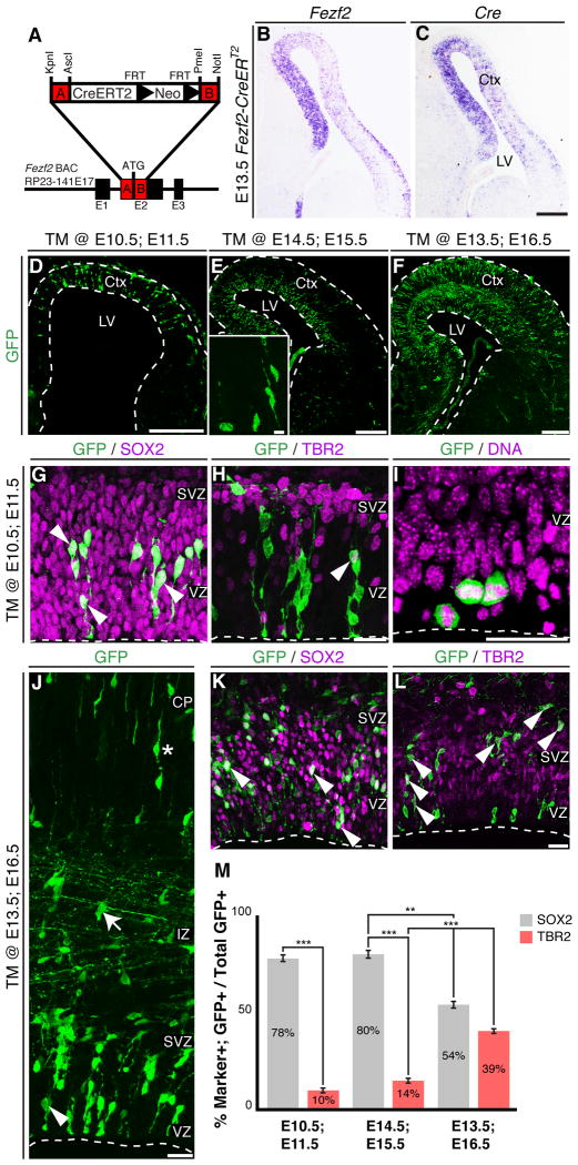
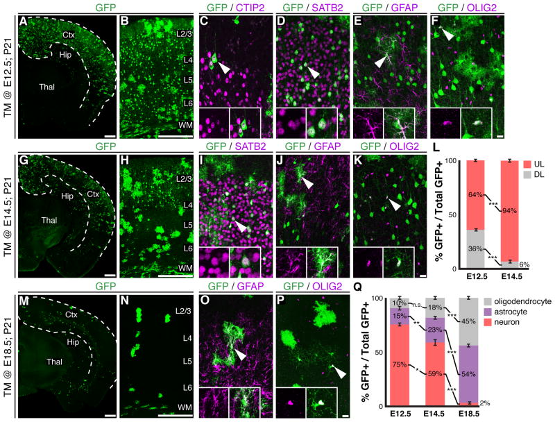
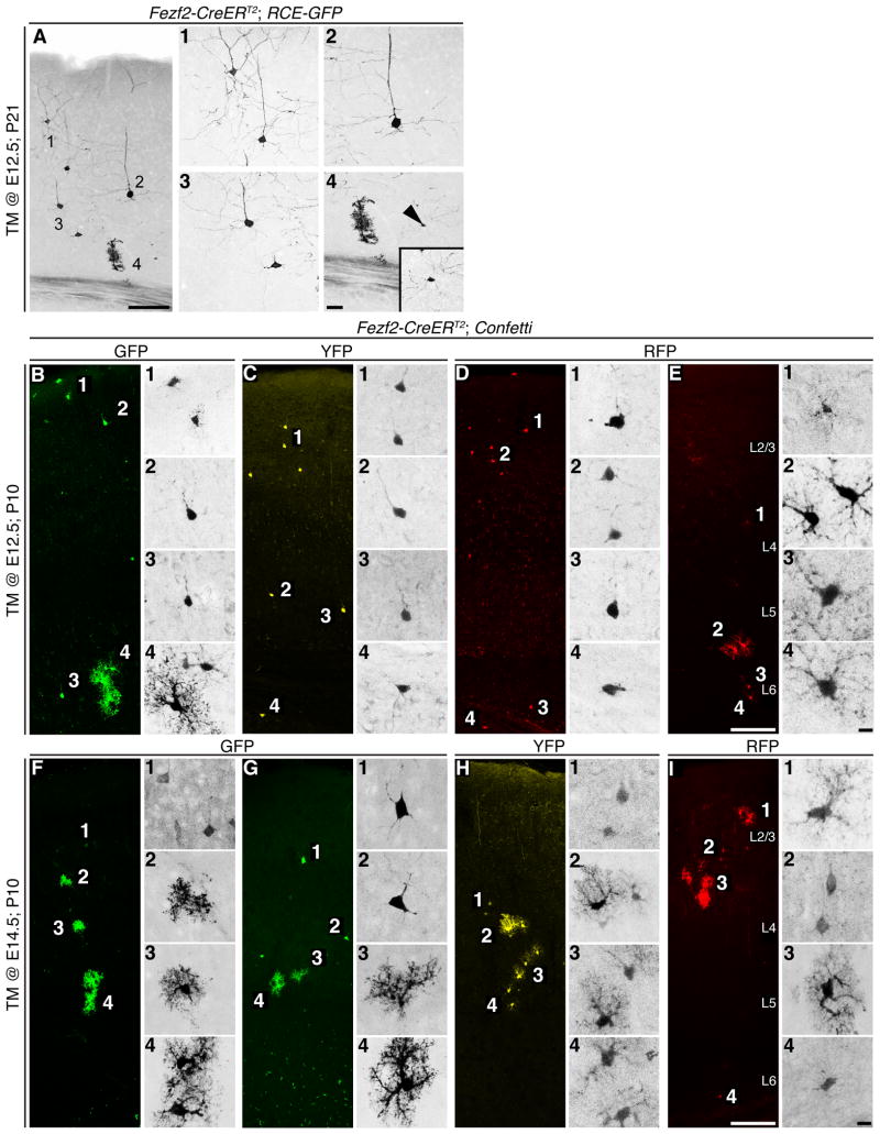
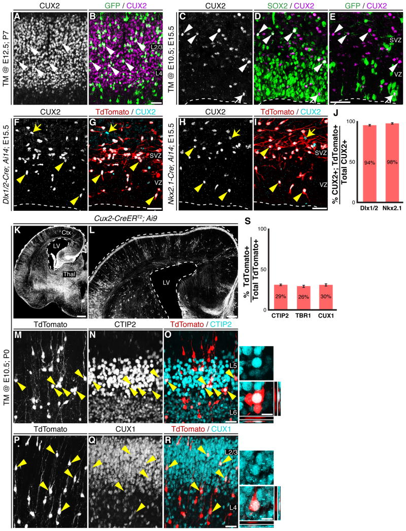
Progenitor cells in the cerebral cortex sequentially generate distinct classes of projection neurons. Recent work suggests the cortex may contain intrinsically fate-restricted progenitors marked by expression of Cux2. However, the heterogeneity of the neocortical ventricular zone as well as the contribution of lineage-restricted progenitors to the overall cortical neurogenic program remains unclear. Here, we utilize in vivo genetic fate mapping to demonstrate that Fezf2-expressing radial glial cells (RGCs) exist throughout cortical development and sequentially generate all major projection neuron subtypes and glia. Moreover, we show that the vast majority of CUX2⁺ cells in the VZ and SVZ are migrating interneurons derived from the subcortical telencephalon. Examination of the embryonic cortical progenitor population demonstrates that Cux2⁺ RGCs generate both deep- and upper-layer projection neurons. These results identify Fezf2⁺ radial glial cells as a multipotent neocortical progenitor and suggest that the existence, and molecular identity, of laminar-fate-restricted RGCs awaits further investigation.


Figures

Figure 1
Figure 1
Fezf2-expressing progenitors are RGCs. (A) Strategy for generation of Fezf2-CreERT2 mice. (B-C) In situ hybridization for Fezf2 (B) and Cre (C) at E13.5. (D-F) Low magnification images of GFP+ cells in the cortex of Fezf2-CreERT2; RCE-GFP mice following CRE-mediated recombination. (G-H) 24 hours after tamoxifen induction, most GFP+ cells expressed SOX2 (78 ± 3%) (G), and few cells expressed TBR2 (10 ± 2%) (H). (I) GFP+ cells dividing at the ventricular surface. (J-L) In TM @ E13.5; E16.5 brains, Fezf2+ RGCs gave rise to basal progenitors. (J) GFP was expressed in both VZ and SVZ progenitors (arrowhead), migrating neurons in the intermediate zone (arrow), and cortical neurons (asterisk). (K) GFP+ cells expressed SOX2 (54% ± 3) and showed typical RGC morphology. (L) Many GFP+ cells expressed TBR2 (39% ± 2). (M) Quantification of the percentages of GFP+SOX2+ RGCs and GFP+TBR2+ basal progenitors among all the GFP+ cells ± SEM. * P < 0.05, ** P < 0.005, *** P < 0.0001. CP, cortical plate; Ctx, cerebral cortex; IZ, intermediate zone; LV, lateral ventricle; SVZ, sub-ventricular zone. Thal, thalamus; TM; tamoxifen; VZ, ventricular zone. Scale bars: (C-F) 250 µm, (E insert) 10 µm, (H, I, J, L) 25 µm. See also Figure S1.
Figure 2
Figure 2
Fezf2+ RGCs sequentially generate projection neurons and glia. (A-F) Immunohistochemical analysis of TM @ E12.5; P21 brains. (A, B) GFP+ cells were present throughout the cortex. GFP+ cells expressed CTIP2 (C), SATB2 (D), GFAP (E) or OLIG2 (F). (G-K) Immunohistochemistry on brain sections from TM @ E14.5; P21 mice. (G, H) GFP+ neurons were mostly in layers 2-4. GFP+ cells expressed SATB2 (I), GFAP (J), or OLIG2 (K). (L) Percentage of GFP+ neurons in deep and upper layers ± SEM. (M-P) Immunohistochemical analysis of brains from TM @ E18.5; P21 mice. GFP+ cells included astrocytes (O) and oligodendrocytes (P). (Q) Percentages of GFP+ cells that were neurons, astrocytes or oligodendrocytes ± SEM. * P < 0.05, ** P < 0.005, *** P < 0.0001. Ctx, cerebral cortex; Hip, hippocampus; Thal, thalamus; TM, tamoxifen; WM, white matter. Scale bars: (A, G, M) 500 µm, (B, H, N) 250 µm, (F, K, P) 25 µm. See also Figure S2.
Figure 3
Figure 3
Clonal analysis of Fezf2+ RGCs. (A) GFP+ clone from a P21 Fezf2-CreERT2; RCE-GFP brain that received TM at E12.5, indicating that early Fezf2+ RGCs generate deep- and upper-layer neurons, astrocytes and oligodendrocytes. (B-I) Fezf2-CreERT2; Confetti mice enabled clonal anaylys of RGCs based upon flourescent protein expression. (B-D) Eamples of TM @ E12.5; P10 brains demonstrating that clones included both deep- and upper-layer neurons and glia. (E) We occasionally observed clones that contained only glia. (F-I) TM @ E14.5; P10 brains contained clones with only upper-layer neruons and glia. TM, tamoxifen. Scale Bars: (A, E, I) 100 µm, (A4) 25 µm, (E4, I4) 10 µm.
Figure 4
Figure 4
CUX2+ cells in VZ/SVZ are migrating interneurons and Cux2-Cre/CreER T2 labeled RGCs generate both deep- and upper-layer projection neurons. (A-B) Fezf2+ RGCs generated CUX2+ upper-layer neurons. (C-E) Immunohistochemical analysis of TM @ E10.5; E15.5 Fezf2-CreERT2; RCE-GFP brains. Few CUX2+ cells in the SVZ expressed SOX2 and these cells re located at the VZ/SVZ boundary (C-D). (E) Rare CUX2+GFP+ cell in the VZ/SVZ. These CUX2+GFP+ cells did not express SOX2 (D-E). The arrowheads in C-E point to the CUX2+SOX2+ cells in VZ/SVZ, the arrows point to a rare GFP+CUX2+ cell. (F-J) Dlx1/2-Cre; Ai14 and Nkx2.1-Cre; Ai14 mice revealed that the majority of CUX2+ cells in the E15.5 neocotical VZ/SVZ were interneurons generated from Dlx1/2 (F-G) or Nkx2.1 (H-I) lineages. (J) Quantification of the percentage of CUX2+TdTomato+ cells among all CUX2+ cells in the VZ/SVZ ± SEM. (K-L) TM @ E10.5; P0 Cux2-CreERT2; Ai9 brains contained TdTomato+ cells in all cortical layers. (M-O) TdTomato+ cells in deep-layers expressed CTIP2. (P-R) Upper-layer TdTomato+ cells expressed CUX1. (S) Percentage of TdTomato+ cells in layers 2-6 that expressed CTIP2, TBR1 or CUX1 ± SEM. CP, cortical plate; IZ, intermediate zone; SVZ, subventricular zone; TM, tamoxifen; VZ, ventricular zone. Scale Bars: (B, E, G, I, O, R) 25 µm, (K) 500 µm, (L) 200 µm, (close-ups from O, R) 10 µm. See also Figures S3 and S4.

Comment in

References

    1. Betancourt J, Katzman S, Chen B. Nuclear factor one b regulates neural stem cell differentiation and axonal projection of corticofugal neurons. The Journal of comparative neurology 2013 - PMC - PubMed
    1. Chen B, Schaevitz LR, McConnell SK. Fezl regulates the differentiation and axon targeting of layer 5 subcortical projection neurons in cerebral cortex. Proceedings of the National Academy of Sciences of the United States of America. 2005a;102:17184–17189. - PMC - PubMed
    1. Chen B, Wang SS, Hattox AM, Rayburn H, Nelson SB, McConnell SK. The Fezf2-Ctip2 genetic pathway regulates the fate choice of subcortical projection neurons in the developing cerebral cortex. Proceedings of the National Academy of Sciences of the United States of America. 2008;105:11382–11387. - PMC - PubMed
    1. Chen JG, Rasin MR, Kwan KY, Sestan N. Zfp312 is required for subcortical axonal projections and dendritic morphology of deep-layer pyramidal neurons of the cerebral cortex. Proceedings of the National Academy of Sciences of the United States of America. 2005b;102:17792–17797. - PMC - PubMed
    1. Cobos I, Long JE, Thwin MT, Rubenstein JL. Cellular patterns of transcription factor expression in developing cortical interneurons. Cerebral cortex. 2006;16(Suppl 1):i82–88. - PubMed

Publication types

MeSH terms