

Risk of scar in the comparison of age-related macular degeneration treatments trials
- PMID: 24314839
- PMCID: PMC3943618
- DOI: 10.1016/j.ophtha.2013.10.019
Risk of scar in the comparison of age-related macular degeneration treatments trials
Abstract
Objective: To describe risk factors for scar in eyes treated with ranibizumab or bevacizumab for neovascular age-related macular degeneration (AMD).
Design: Prospective cohort study within a randomized clinical trial.
Participants: Patients with no scar on color fundus photography (CFP) or fluorescein angiography (FA) at enrollment in the Comparison of Age-related Macular Degeneration Treatments Trials (CATT).
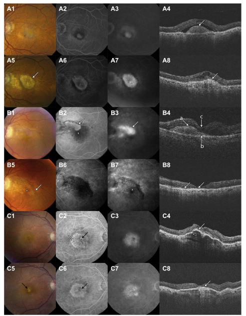
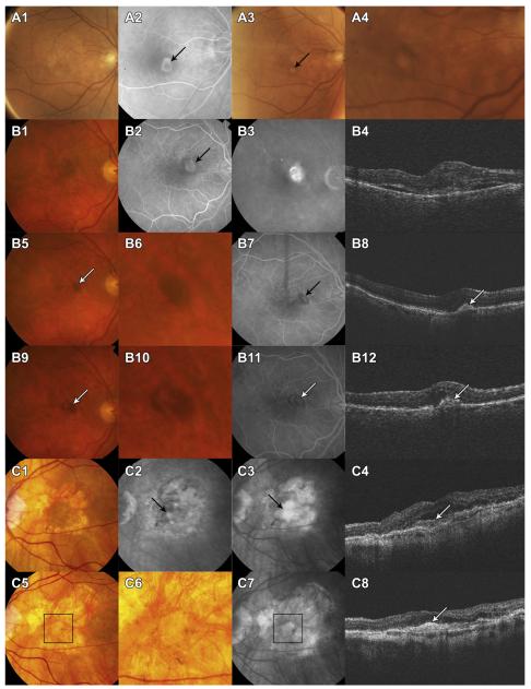
Methods: Eyes were assigned to ranibizumab or bevacizumab treatment and to 1 of 3 dosing regimens for 2 years. Masked readers assessed CFP and FA. Baseline demographic characteristics, visual acuity, morphologic features on photography and optical coherence tomography (OCT), and genotypes associated with AMD risk were evaluated as risk factors using adjusted hazard ratios (aHRs) and associated 95% confidence intervals (CIs). Scars were classified as fibrotic with well-demarcated elevated mounds of yellowish white tissue or nonfibrotic with discrete flat areas of hyperpigmentation with varying amounts of central depigmentation.
Main outcome measures: Scar formation.
Results: Scar developed in 480 of 1059 eyes (45.3%) by 2 years. Baseline characteristics associated with greater risk of scarring were predominantly classic choroidal neovascularization (CNV) (aHR, 3.1; CI, 2.4-3.9) versus occult CNV, blocked fluorescence (aHR, 1.4; CI, 1.1-1.8), foveal retinal thickness >212 μm (aHR, 2.4; CI, 1.7-3.6) versus <120 μm, foveal subretinal tissue complex thickness >275 μm (aHR, 2.4; CI, 1.7-3.6) versus ≤75 μm, foveal subretinal fluid (aHR, 1.5; CI, 1.1-2.0) versus no subretinal fluid, and subretinal hyperreflective material (SHRM) (aHR, 1.7; CI, 1.3-2.3) versus no SHRM. Eyes with elevation of the retinal pigment epithelium had lower risk (aHR, 0.6; CI, 0.5-0.8) versus no elevation. Drug, dosing regimen, and genotype had no statistically significant association with scarring. Fibrotic scars developed in 24.7% of eyes, and nonfibrotic scars developed in 20.6% of eyes. Baseline risk factors for the scar types were similar except that eyes with larger lesion size or visual acuity <20/40 were more likely to develop fibrotic scars.
Conclusions: Approximately half of eyes enrolled in CATT developed scar by 2 years. Eyes with classic neovascularization, a thicker retina, and more fluid or material under the foveal center of the retina are more likely to develop scar.
Trial registration: ClinicalTrials.gov NCT00593450.
Copyright © 2014 American Academy of Ophthalmology. Published by Elsevier Inc. All rights reserved.
Figures
References
-
- Bressler NM, Frost LA, Bressler SB, et al. Natural course of poorly defined choroidal neovascularization associated with macular degeneration. Arch Ophthalmol. 1988;106:1537–42. - PubMed
-
- Wong TY, Chakravarthy U, Klein R, et al. The natural history and prognosis of neovascular age-related macular degeneration: a systematic review of the literature and meta-analysis. Ophthalmology. 2008;115:116–26. - PubMed
-
- Pauleikhoff D. Neovascular age-related macular degeneration: natural history and treatment outcomes. Retina. 2005;25:1065–84. - PubMed
-
- Sivaprasad S, Saleh GM, Jackson H. Does lesion size determine the success rate of photodynamic therapy for age-related macular degeneration? Eye (Lond) 2006;20:43–5. - PubMed
Publication types
MeSH terms
Substances
Associated data
Grants and funding
LinkOut - more resources
Full Text Sources
Other Literature Sources
Medical
Miscellaneous
