

Lateral rectus superior compartment palsy
- PMID: 24315033
- PMCID: PMC4000173
- DOI: 10.1016/j.ajo.2013.09.027
Lateral rectus superior compartment palsy
Abstract
Purpose: To employ magnetic resonance imaging (MRI) to seek evidence of compartmental lateral rectus atrophy consistent with a lesion involving selective denervation of only 1 of the 2 neuromuscular compartments of the lateral rectus.
Design: Prospective observational case-control series.
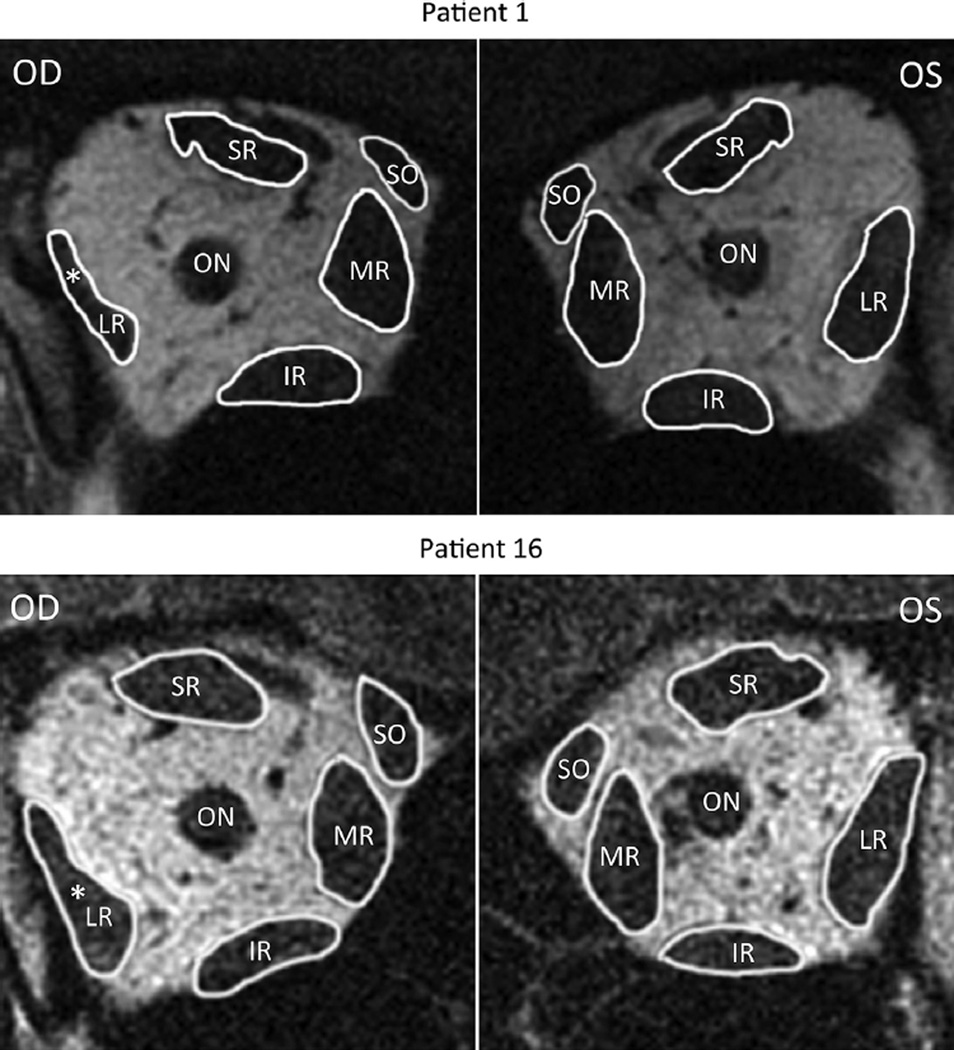
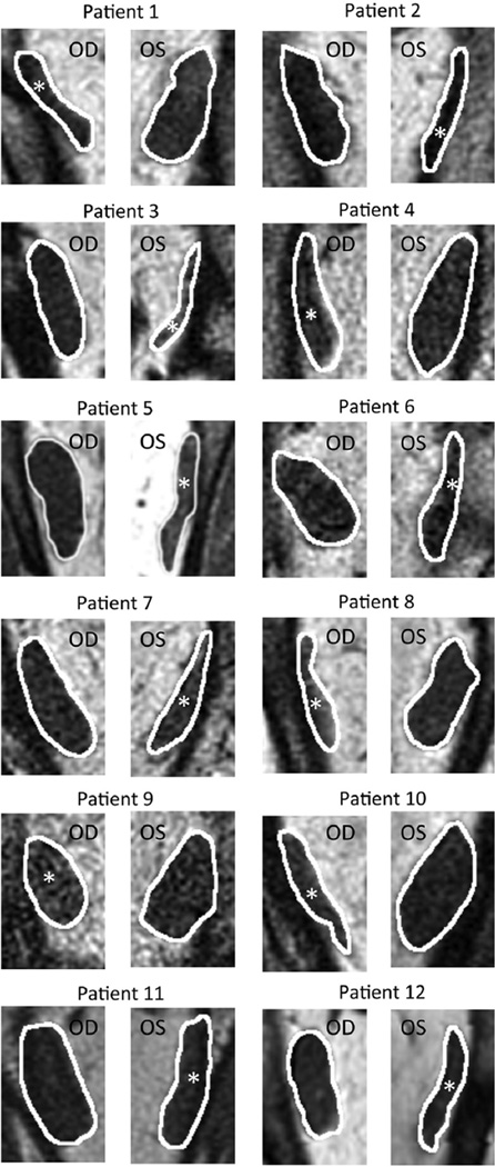
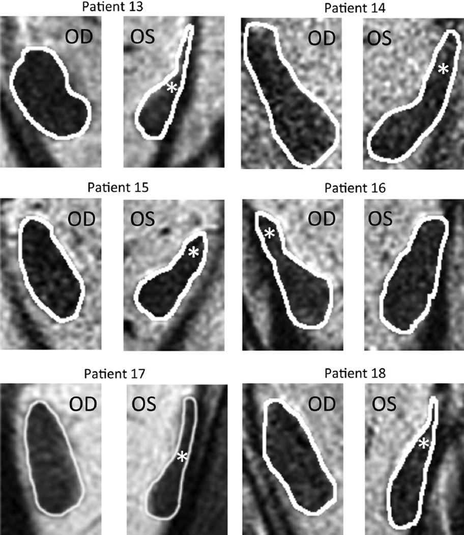
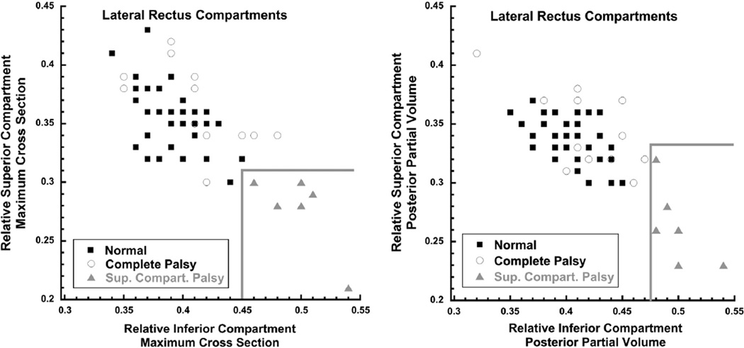
Methods: At a single institution, surface coil coronal MRI was obtained at 312 μm resolution in quasi-coronal planes 2 mm thick throughout the orbit in 20 normal volunteers and 18 patients with unilateral lateral rectus palsy fixated monocularly on a target placed in central gaze. Maximum cross sections and posterior volumes of the superior and inferior lateral rectus compartments were computed and correlated with clinical findings.
Results: Twelve patients with lateral rectus palsy demonstrated symmetric, highly significant 40% reductions in maximum cross sections and 50% reductions in posterior volumes from normal for both compartments (P < 10(-6) for all comparisons). Six patients with lateral rectus palsy had similar significant but asymmetric reductions in those measures only for the superior compartment of the affected lateral rectus (P < 10(-4) for all comparisons), with insignificant 20%-30% reductions for the inferior compartment (P > 0.2 for all comparisons).
Conclusions: A subset of patients with clinical lateral rectus palsy may have palsy limited to the superior compartment. Paralytic esotropia may be caused by lateral rectus superior compartment palsy despite an intact lateral rectus inferior compartment. This finding is consistent with evidence supporting independent innervation of the 2 lateral rectus neuromuscular compartments.
Copyright © 2014. Published by Elsevier Inc.
Conflict of interest statement
ALL AUTHORS HAVE COMPLETED AND SUBMITTED THE ICMJE FORM FOR DISCLOSURE OF POTENTIAL CONFLICTS OF INTEREST. None of the authors have any financial disclosures to report. The authors have no proprietary interests in the surface coils.
Figures

Comment in
-
Lateral Rectus Superior Compartment Palsy.Am J Ophthalmol. 2015 Jul;160(1):205-6. doi: 10.1016/j.ajo.2015.03.026. Am J Ophthalmol. 2015. PMID: 26054471 No abstract available.
-
Reply: To PMID 24315033.Am J Ophthalmol. 2015 Jul;160(1):206-7. doi: 10.1016/j.ajo.2015.03.025. Am J Ophthalmol. 2015. PMID: 26054472 No abstract available.
Similar articles
-
Extraocular Muscle Compartments in Superior Oblique Palsy.Invest Ophthalmol Vis Sci. 2016 Oct 1;57(13):5535-5540. doi: 10.1167/iovs.16-20172. Invest Ophthalmol Vis Sci. 2016. PMID: 27768791 Free PMC article.
-
Comparison of orbital magnetic resonance imaging in duane syndrome and abducens palsy.Am J Ophthalmol. 2006 Nov;142(5):827-34. doi: 10.1016/j.ajo.2006.06.012. Epub 2006 Sep 20. Am J Ophthalmol. 2006. PMID: 16989758 Free PMC article.
-
Lateral Rectus Superior Compartment Palsy.Am J Ophthalmol. 2015 Jul;160(1):205-6. doi: 10.1016/j.ajo.2015.03.026. Am J Ophthalmol. 2015. PMID: 26054471 No abstract available.
-
Vertical rectus transposition procedures for lateral rectus palsy: A systematic review.Indian J Ophthalmol. 2019 Nov;67(11):1793-1799. doi: 10.4103/ijo.IJO_1841_18. Indian J Ophthalmol. 2019. PMID: 31638036 Free PMC article.
-
Compartmental Strabismus.J Binocul Vis Ocul Motil. 2020 Jul-Sep;70(3):71-78. doi: 10.1080/2576117X.2020.1760683. Epub 2020 May 28. J Binocul Vis Ocul Motil. 2020. PMID: 32463321 Review.
Cited by
-
Recent advances clarifying the etiologies of strabismus.J Neuroophthalmol. 2015 Jun;35(2):185-93. doi: 10.1097/WNO.0000000000000228. J Neuroophthalmol. 2015. PMID: 25724009 Free PMC article. Review.
-
Hypertropia in unilateral isolated abducens palsy.J AAPOS. 2014 Jun;18(3):235-40. doi: 10.1016/j.jaapos.2014.01.017. J AAPOS. 2014. PMID: 24924275 Free PMC article.
-
Extraocular Muscle Compartments in Superior Oblique Palsy.Invest Ophthalmol Vis Sci. 2016 Oct 1;57(13):5535-5540. doi: 10.1167/iovs.16-20172. Invest Ophthalmol Vis Sci. 2016. PMID: 27768791 Free PMC article.
-
Magnetic Resonance Imaging of the Globe-Tendon Interface for Extraocular Muscles: Is There an "Arc of Contact"?Am J Ophthalmol. 2018 Oct;194:170-181. doi: 10.1016/j.ajo.2018.07.002. Epub 2018 Jul 19. Am J Ophthalmol. 2018. PMID: 30030978 Free PMC article.
-
Functional anatomy of extraocular muscles during human vergence compensation of horizontal heterophoria.J Neurophysiol. 2019 Jul 1;122(1):105-117. doi: 10.1152/jn.00152.2019. Epub 2019 May 1. J Neurophysiol. 2019. PMID: 31042451 Free PMC article.
References
-
- von Noorden G, Chu MW. Surgical treatment options in cyclotropia. J Pediatr Ophthalmol Strabismus. 1990;27(6):291–293. - PubMed
Publication types
MeSH terms
Grants and funding
LinkOut - more resources
Full Text Sources
Other Literature Sources
Medical
