. 2014 Jan:13:10-21.
doi: 10.1016/j.dnarep.2013.10.011. Epub 2013 Dec 4.

Mammalian MutY homolog (MYH or MUTYH) protects cells from oxidative DNA damage

Affiliations

Mammalian MutY homolog (MYH or MUTYH) protects cells from oxidative DNA damage

Bor-Jang Hwang et al. DNA Repair (Amst). 2014 Jan.

Abstract

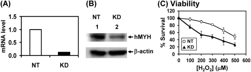
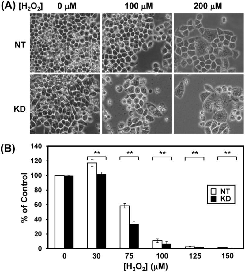
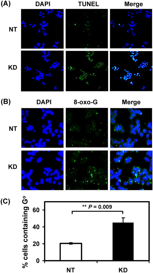
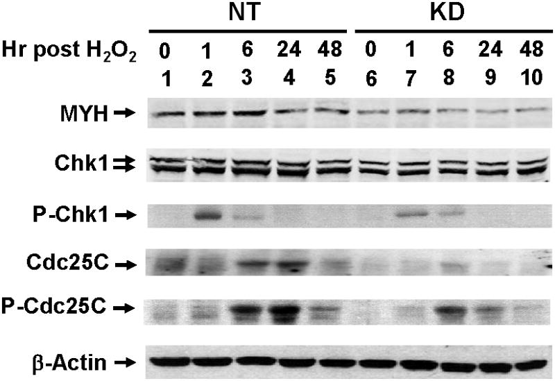
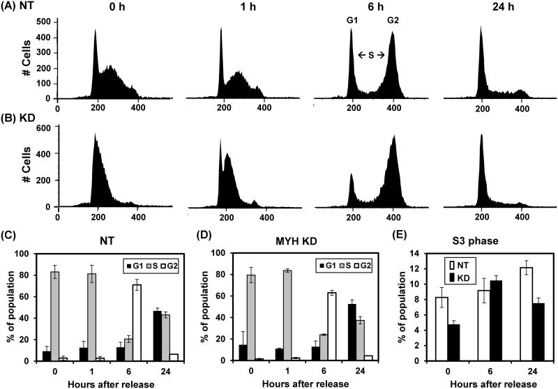
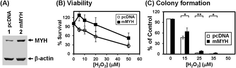
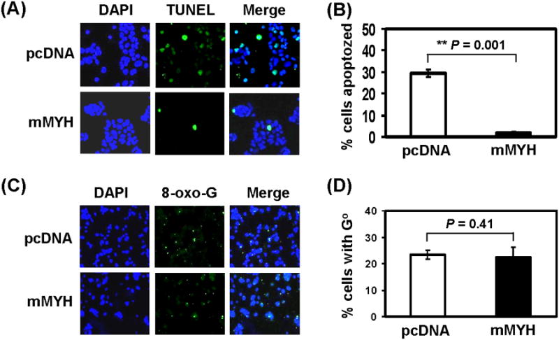
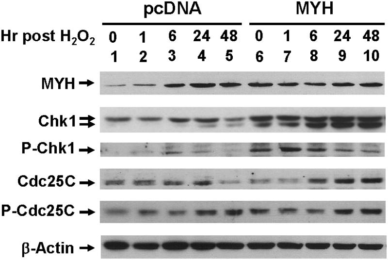
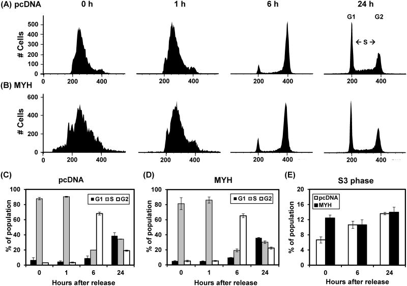
MutY DNA glycosylase homologs (MYH or MUTYH) reduce G:C to T:A mutations by removing misincorporated adenines or 2-hydroxyadenines paired with guanine or 8-oxo-7,8-dihydroguanine (8-oxo-G). Mutations in the human MYH (hMYH) gene are associated with the colorectal cancer predisposition syndrome MYH-associated polyposis. To examine the function of MYH in human cells, we regulated MYH gene expression by knockdown or overproduction. MYH knockdown human HeLa cells are more sensitive to the killing effects of H2O2 than the control cells. In addition, hMYH knockdown cells have altered cell morphology, display enhanced susceptibility to apoptosis, and have altered DNA signaling activation in response to oxidative stress. The cell cycle progression of hMYH knockdown cells is also different from that of the control cells following oxidative stress. Moreover, hMYH knockdown cells contain higher levels of 8-oxo-G lesions than the control cells following H2O2 treatment. Although MYH does not directly remove 8-oxo-G, MYH may generate favorable substrates for other repair enzymes. Overexpression of mouse Myh (mMyh) in human mismatch repair defective HCT15 cells makes the cells more resistant to killing and refractory to apoptosis by oxidative stress than the cells transfected with vector. In conclusion, MYH is a vital DNA repair enzyme that protects cells from oxidative DNA damage and is critical for a proper cellular response to DNA damage.

Keywords: Base excision repair; DNA damage response; DNA repair; MutY homolog; Oxidative stress.


Conflict of interest statement

Conflict of interest statement: The authors declare that there is no conflict of interest.

Figures

Fig. 1
Fig. 1
HeLa cells with hMYH-knockdown are more sensitive to H2O2 than control. (A) mRNA level of hMYH is reduced to 12% in knockdown (KD) cells as compared to non-target (NT) control cells. HeLa cells were stably transfected with lentivirus containing shRNA against hMYH or control shRNA. mRNA levels were determined by RT-qPCR. (B) The protein level of hMYH is reduced to 30% in knockdown cells as compared to control cells. Extracts were prepared from control and knockdown cells and Western blotting were analyzed with hMYH and β-actin antibodies. (C) Sensitivity to H2O2 of MYH KD cells. Both non-target and MYH knockdown HeLa cells were treated with H2O2 at indicated concentrations for 1 hr. The survived cells were detected after 24 hrs. Survival differences are statistical significant between two cell lines at 100-500 μM H2O2.
Fig. 2
Fig. 2
HeLa cells with hMYH-knockdown have altered cell morphology and poor survival rate than control cells after H2O2 treatment. HeLa cells were stably transfected with lentivirus containing shRNA against hMYH or non-target (NT) shRNA. (A) Morphological changes of MYH KD cells after H2O2 treatment. MYH knockdown HeLa cells were treated with 0, 100 and 200 μM H2O2 for 1 hour. Cells were imaged under microscope after 6 days of treatment. Fewer cell number count and more cells undergo morphological changes were observed in H2O2-treated hMYH KD cells as compared to control cells. (B) Colony formation of MYH KD cells after H2O2 treatment. Cells were treated with 30-150 μM H2O2 for 1 h or left untreated (control). Plates were incubated for 10 days. Quantitative analyses of the relative colony formation of NT (white bar) and MYH-ND (black bars) from three experiments. The percentage (%) is calculated from the ratios of H2O2 treated over untreated samples. The error bars reported are the standard deviations of the averages and P-value was calculated by Student's t-test. Two stars indicate P < 0.05.
Fig. 3
Fig. 3
HeLa cells with hMYH knockdown are more sensitive to apoptosis and contain higher level of 8-oxoG than control cells after H2O2 treatment. HeLa cells were stably transfected with lentivirus containing shRNA against hMYH or non-target (NT) shRNA. (A) Cells were treated with 150 μM H2O2 for 1 hr, replaced with fresh media for one day, and subjected to DAPI staining and TUNEL assay. Both were merged in the third column. (B) Cells were treated with 150 μM H2O2 for 1 hr and subjected to immuno-florescent staining with anti-8-oxo-dG antibody. Nuclear DNA was counterstained with DAPI. Upper panel, cells with control NT shRNA. Lower panel was from cells with hMYH knockdown. (C) Quantitative analyses of GO accumulation of NT (white bar) and MYH-ND (black bars) cells from three experiments. The percentage (%) of cells containing GO is presented after H2O2 treatment. The error bars reported are the standard deviations of the averages and P-value shown was calculated by Student's t-test.
Fig. 4
Fig. 4
DNA damage signaling in hMYH-knockdown HeLa cells after H2O2 treatment. HeLa cells were stably transfected with lentivirus containing shRNA against hMYH or non-target (NT) shRNA. Cells were treated with 150 μM H2O2 for 1 hr and then recovered for different time intervals as indicated. Cell extracts were prepared for Western blotting analysis with antibodies against different proteins. P-Chk1 and P-Cdc25C are Ser 317-phosphorylated Chk1 and Ser 216-phosphorylated Cdc25C, respectively.
Fig. 5
Fig. 5
Cell cycle progression in control and MYH knockdown HeLa cells after exposure to H2O2. HeLa cells were stably transfected with lentivirus containing shRNA against hMYH (KD) or non-target (NT) shRNA. Cells were synchronized at S phase by double thymidine block, treated with 75 μM H2O2 for one hour, and then collected for cell cycle analysis. 0 hr reflects the time of H2O2 treatment and removal of thymidine. (A) The FACS profiles of NT control cells. (B) The FACS profiles of MYH knockdown cells. (C) and (D), Quantitative evaluations of (A) and (B), respectively. The panels show the percentage of population in G1 (black bars), S (grey bars) and G2 (open bars) phases of the cell cycle. (E) Quantitative analyses of late S-phase (S3) population in both NT and MYH KD cells.
Fig. 6
Fig. 6
HCT15 (MSH6-defective) human cells expressing exogenous mMYH is more resistant to hydrogen peroxide. (A) Mouse MYH protein was over-expressed in HCT15 cells. Cells were transfected with pcDNA (vector) or mMYH. Extracts were prepared for Western blotting with hMYH and actin antibodies. Both endogenous hMYH and expressed mMYH were detected. (B) Sensitivity to H2O2 of MYH overproducing cells. Both control and MYH overproducing cells were treated with H2O2 at indicated concentrations for 1 hr. The survived cells were detected after 24 hrs. Survival differences are statistical significant between two cell lines at 5, 10, and 20 μM H2O2. (C) Quantitative analyses of the relative colony formation in cells containing vector (white bar) or mMYH (black bar) from three experiments. The percentage (%) is calculated from the ratios of H2O2 treated over untreated samples. The error bars reported are the standard deviations of the averages and P-value was calculated by Student's t-test. One star indicates P < 0.1 and two stars indicate P < 0.05.
Fig. 7
Fig. 7
HCT15 (MSH6-defective) human cells expressing exogenous mMYH are more refractory to apoptosis than control cells, but contain the same levels of 8-oxo-G lesions as control cells expressing only endogenous hMYH after peroxide treatment. (A) Cells were stably transfected with pcDNA or mMYH and then treated with 0 or 25 μM H2O2 for 1 hr, recovered in fresh media for one day, and subjected to TUNEL analysis and DAPI staining. (B) Quantitative analyses of apoptosis in cells containing vector (white bar) or mMYH (black bar) from three experiments. The percentage (%) of apoptozed cells is presented after H2O2 treatment. The error bars reported are the standard deviations of the averages and P-value shown was calculated by Student's t-test (C) Cells similar as in (A) were treated with 35 μM H2O2 for 1 hr and subjected to immuno-florescent staining with 8-oxo-dG antibody and DAPI staining. (D) Quantitative analyses of GO accumulation in cells containing vector (white bar) or mMYH (black bar) from three experiments. The percentage (%) of cells containing GO is presented after H2O2 treatment. The error bars reported are the standard deviations of the averages and P-value shown was calculated by Student's t-test.
Fig. 8
Fig. 8
DNA damage signaling in mMYH-overproducing HCT15 cells after H2O2 treatment. Cells transfected with pcDNA or mMYH were treated with 25 μM H2O2 for 1 hr, and then recovered for different time intervals as indicated. Cell extracts were subjected to Western blotting analysis. P-Chk1 and P-Cdc25C are Ser 317-phosphorylated Chk1 and Ser 216-phosphorylated Cdc25C, respectively.
Fig. 9
Fig. 9
Cell cycle progression in control and MYH overproducing HCT15 cells after exposure to H2O2. Cells stably transfected with pcDNA or mMYH were synchronized at S phase by double thymidine block, treated with 25 μM H2O2 for one hour, and then collected for cell cycle analysis. (A) The FACS profiles of control cells. 0 hr reflects the time of H2O2 treatment and removal of thymidine. (B) The FACS profiles of MYH overproducing cells. (C) and (D), Quantitative evaluations of (A) and (B), respectively. The panels show the percentage of population in G1 (black bars), S (grey bars) and G2 (open bars) phases of the cell cycle. (E) Quantitative analyses of late S-phase (S3) population in both control and MYH overproducing cells.

References

    1. Friedberg EC, Walker GC, Siede W, Wood RD, Schultz RA, Ellenberger T. DNA Repair and Mutagenesis. ASM Press; Washington, D.C.: 2005.
    1. Hoeijmakers JH. Genome maintenance mechanisms for preventing cancer. Nature. 2001;411:366–374. - PubMed
    1. Krokan HE, Nilsen H, Skorpen F, Otterlei M, Slupphaug G. Base excision repair of DNA in mammalian cells. FEBS Lett. 2000;476:73–77. - PubMed
    1. Mol CD, Parikh SS, Putnam CD, Lo TP, Tainer JA. DNA repair mechanisms for the recognition and removal of damaged DNA bases. Annu REv Biophys Biomol Struct. 1999;28:101–128. - PubMed
    1. Avkin S, Livneh Z. Efficiency, specificity and DNA polymerase-dependence of translesion replication across the oxidative DNA lesion 8-oxoguanine in human cells. Mutat Res. 2002;510:81–90. - PubMed

Publication types

MeSH terms