. 2013 Dec 5;13(6):676-90.
doi: 10.1016/j.stem.2013.10.008.

The THO complex regulates pluripotency gene mRNA export and controls embryonic stem cell self-renewal and somatic cell reprogramming

Affiliations

The THO complex regulates pluripotency gene mRNA export and controls embryonic stem cell self-renewal and somatic cell reprogramming

Li Wang et al. Cell Stem Cell. .

Abstract

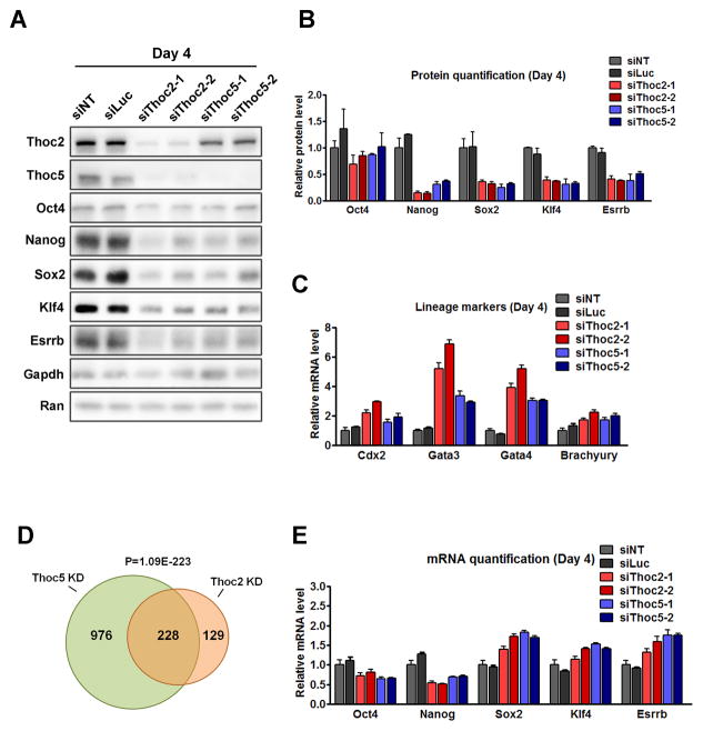
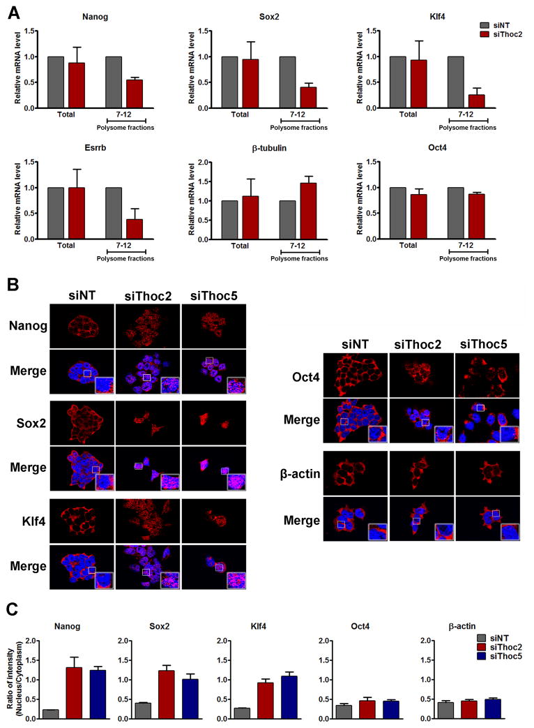
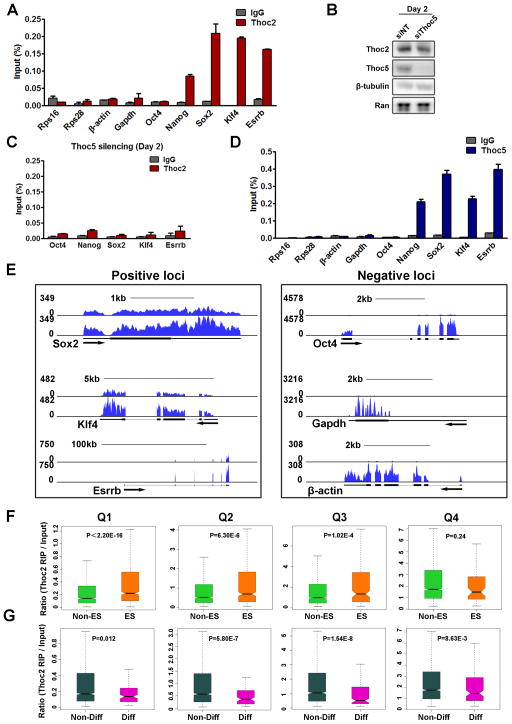
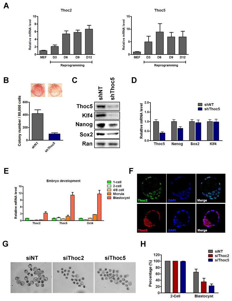
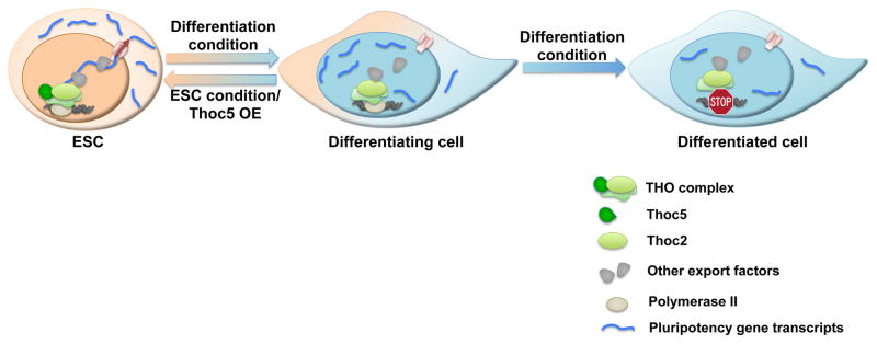
Embryonic stem cell (ESC) self-renewal and differentiation are governed by a broad-ranging regulatory network. Although the transcriptional regulatory mechanisms involved have been investigated extensively, posttranscriptional regulation is still poorly understood. Here we describe a critical role of the THO complex in ESC self-renewal and differentiation. We show that THO preferentially interacts with pluripotency gene transcripts through Thoc5 and is required for self-renewal at least in part by regulating their export and expression. During differentiation, THO loses its interaction with those transcripts due to reduced Thoc5 expression, leading to decreased expression of pluripotency proteins that facilitates exit from self-renewal. THO is also important for the establishment of pluripotency, because its depletion inhibits somatic cell reprogramming and blastocyst development. Together, our data indicate that THO regulates pluripotency gene mRNA export to control ESC self-renewal and differentiation, and therefore uncover a role for this aspect of posttranscriptional regulation in stem cell fate specification.


Figures

Figure 1
Figure 1. Thoc2 and Thoc5 expression correlates with the pluripotent state of ESCs
(A) Expression of Thoc2 and Thoc5 during ESC differentiation. Thoc2, Thoc5, and Oct4 expression was determined by RT-qPCR at the indicated time points during ESC differentiation. Differentiation was induced by LIF-withdrawal, retinoic acid (RA) treatment, or embryoid body (EB) formation. Expression was normalized to day-0 by β-actin and plotted as mean ± SEM. (B) Oct4GiP reporter assay: the Oct4GiP reporter ESCs were transfected with non-targeting (NT), luciferase (Luc), Thoc2, or Thoc5 siRNAs, and the reporter activity was determined by fluorescence-activated cell sorting (FACS) analysis 96 hours after transfection. The percentage of differentiation was determined by the percentage of GFP-negative cells and plotted as mean ± SEM. (C) ESC morphology after Thoc2 and Thoc5 silencing. ESCs were transfected with the indicated siRNAs, and cellular morphology was examined 96 hours after transfection. See also Figure S1, S2.
Figure 2
Figure 2. Thoc2 or Thoc5 depletion results in loss of ESC self-renewal
(A) Expression of the pluripotency genes after Thoc2 and Thoc5 silencing. Expression was determined by western blot 96 hours after siRNA transfections. GAPDH and RAN were used as loading controls. (B) Quantitation of the pluripotency protein expression based on western blot. Expression was normalized by GAPDH and compared to the NT-siRNA transfected cells, and plotted as mean ± SEM from four independent experiments. (C) Lineage marker expression after Thoc2 and Thoc5 silencing. Expression was determined by RT-qPCR 96 hours after siRNA transfection. Values are plotted as mean ± SEM from three independent experiments. (D) Venn diagram of genes differentially expressed 96 hours after Thoc2 or Thoc5 siRNA transfection based on microarray. (E) Pluripotency gene mRNA levels were determined by RT-qPCR 96 hours after siRNA transfection. Values are plotted as mean ± SEM from three independent experiments. See also Figure S3, Table S1, S2.
Figure 3
Figure 3. Thoc2 or Thoc5 depletion inhibits the export of pluripotency gene mRNAs
(A) Effect of Thoc2 silencing on polysome-associated RNAs. ESCs were transfected with NT or Thoc2 siRNAs, and cells were harvested 96 hours after transfection. Polysome-associated RNAs were purified by sucrose-gradient ultracentrifugation. Relative abundance of mRNAs in total (Total) or polysome-associated (Fraction 7–12) RNAs was determined by RT-qPCRs, and plotted as mean ± SEM from three independent experiments. (B) Effect of Thoc2 or Thoc5 silencing on mRNA export. ESCs were transfected with NT, Thoc2, or Thoc5 siRNAs. RNA Fluorescence in situ hybridization (RNA-FISH) was carried out for Nanog, Sox2, Klf4, Oct4 and β-actin 96 hours after transfection. Red: fluorescence signals from the FISH probes. Blue: DAPI staining of the nuclei. (C) Ratio of the RNA FISH signals in the nucleus versus cytoplasm was quantified by Metamorph from at least 10 images from three independent experiments. Values are plotted as mean ± SEM. See also Figure S4.
Figure 4
Figure 4. Thoc2 preferentially binds to pluripotency gene mRNAs in a Thoc5-dependent manner
(A) Thoc2 interaction with mRNAs. RIP was performed in ESCs with either IgG or Thoc2 antibody. Relative abundance of the immunoprecipitated mRNAs was determined by RT-qPCRs and normalized to Input, and plotted as mean ± SEM from three independent experiments. (B) Thoc2 protein level 48 hours after Thoc5 silencing. (C) Effect of Thoc5 silencing on Thoc2 RIP. ESCs were transfected with Thoc5 siRNAs, and RIP was carried out 48 hours after transfection. Values are plotted as mean ± SEM. (D) Thoc5 interaction with mRNAs. RIP was performed in ESCs. Relative abundance of the immunoprecipitated mRNAs was determined by RT-qPCRs and normalized to Input, and plotted as mean ± SEM from three independent experiments. (E) Thoc2 RIP-sequencing. Thoc2 RIP was performed in ESCs, and the total and Thoc2-interacting RNAs were subjected to high-throughput sequencing. Representative images from the genome browser were shown for the indicated genes. (F–G) Statistical analysis of Thoc2 RIP-sequencing. Genes detected in the sequencing were ranked by their expression level in ESCs and divided into 4 equal groups (Q1–Q4). Thoc2 binding was determined by the ratio of the reads per kilo base per million (RPKM) in Thoc2 RIP/Input, and was compared between non-ES cell genes (Non-ES) and ES cell genes (ES) or non-differentiation (Non-Diff) and differentiation (Diff) genes in each quarter. ES genes were defined as those down-regulated during ESC differentiation, while differentiation genes were defined as those up-regulated (see methods for details). p-values were calculated by the Wilcoxon rank sum test.
Figure 5
Figure 5. Thoc5 regulates the exit from the pluripotent state
(A–B) Expression of Thoc5 and pluripotency genes during ESC differentiation induced by LIF withdrawal. Protein expression was determined by western blot (A). mRNA expression was determined by RT-qPCRs (B). Values were plotted as mean ± SEM from three independent experiments. (C) Thoc2 RIP after LIF withdrawal. RIP was carried out 24 hours after LIF withdrawal. Immunoprecipitated mRNAs were quantitated by RT-qPCRs and normalized to Input, and plotted as mean ± SEM from three independent experiments. (D) Effect of Thoc5 overexpression on Thoc2 interaction with the pluripotency gene mRNAs. RIP was carried out in the GFP (GFPOE) or Thoc5 overexpression (Thoc5OE) ESCs 24 hours after LIF withdrawal. Immunoprecipitated mRNAs were quantitated by RT-qPCRs and normalized to the input, and plotted as mean ± SEM from three independent experiments. *: p < 0.05. (E) Effect of Thoc5 overexpression on pluripotency gene expression. Expression of pluripotency gene proteins in GFP (Ind-GFP) or Thoc5 overexpression ESCs (two different lines Ind-Thoc5-1 and Ind-Thoc5-2) at the indicated time points during LIF withdrawal was determined by western blot. (F–H) Effect of Thoc5 overexpression on ESC self-renewal. (F) GFP (Ind-GFP) or Thoc5 (Ind-Thoc5-1, Ind-Thoc5-2) overexpression ESCs were cultured without LIF for the indicated amount of time, and the different types of colonies formed were counted and plotted as percentage of total. Values are plotted as mean from three independent experiments. (G) GFP (GFPOE) or Thoc5 overexpression (Thoc5OE) ESCs were cultured without LIF for the indicated amount of time. The expression of the differentiation markers was determined by RT-qPCRs and plotted as mean ± SEM from three independent experiments. (H) GFP (GFPOE) or Thoc5 (Thoc5OE) overexpression ESCs were cultured without LIF for 48 hours, and then re-plated at clonal density in ESC culture conditions. The number of ESC colonies formed 8 days later was counted and plotted as mean ± SEM from three independent experiments. Representative images of differentiated and undifferentiated colonies are shown at the top. See also Figure S5, S6.
Figure 6
Figure 6. THO depletion inhibits reprogramming and blastocyst development
(A) Thoc2 and Thoc5 expression during reprogramming as determined by RT-qPCRs. Values are plotted as mean ± SEM from three independent experiments. (B) Effect of Thoc5 KD on reprogramming. MEFs were transduced with NT or Thoc5 shRNA viruses, and then transduced with the OKSM viruses to initiate reprogramming. Number of AP-positive colonies was counted 9 days after the start of the reprogramming, and values are plotted as mean ± SEM from four independent experiments. (C-D) The expression of Thoc5 and pluripotency genes was determined at the end of reprogramming. The protein expression was determined by western blot (C). The mRNA expression was determined by RT-qPCRs (D). Values are plotted as mean ± SEM from three independent experiments. (E) Thoc2 and Thoc5 expression during early embryonic development in vivo. mRNA expression was determined by RT-qPCRs and normalized to the 1-cell stage. Values are plotted as mean ± SEM. (F) Immunofluorescence staining of Thoc2 (green) and Thoc5 (red) in E3.5 blastocysts. DAPI (Blue) was used to stain the cell nuclei. (G–H) Effect of Thoc2 or Thoc5 KD on blastocyst development. (G) 1-cell embryos were injected with NT, Thoc2, or Thoc5 siRNAs and cultured for 4 days ex vivo. Images of the embryos were taken on day-4. (H) The number of normal blastocyst formed on day-4 was counted and normalized to the number of normal 2-cell embryos formed on day-2, and plotted as mean ± SEM from three independent experiments. See also Figure S7.
Figure 7
Figure 7. THO controls pluripotency gene mRNA export to regulate ESC self-renewal and differentiation
In ESCs, THO is necessary for the efficient export of pluripotency gene mRNAs and the maintenance of self-renewal. Upon differentiation or Thoc5 down-regulation, decreased Thoc5 level results in impaired pluripotency gene mRNA export and expression, leading to differentiation. By post-transcriptionally regulating pluripotency gene expression, THO provides an important layer of control to regulate ESC self-renewal and differentiation.

Comment in

References

    1. Boyer LA, Lee TI, Cole MF, Johnstone SE, Levine SS, Zucker JP, Guenther MG, Kumar RM, Murray HL, Jenner RG, et al. Core transcriptional regulatory circuitry in human embryonic stem cells. Cell. 2005;122:947–956. - PMC - PubMed
    1. Buckley SM, Aranda-Orgilles B, Strikoudis A, Apostolou E, Loizou E, Moran-Crusio K, Farnsworth CL, Koller AA, Dasgupta R, Silva JC, et al. Regulation of pluripotency and cellular reprogramming by the ubiquitin-proteasome system. Cell stem cell. 2012;11:783–798. - PMC - PubMed
    1. Chambers I, Silva J, Colby D, Nichols J, Nijmeijer B, Robertson M, Vrana J, Jones K, Grotewold L, Smith A. Nanog safeguards pluripotency and mediates germline development. Nature. 2007;450:1230–1234. - PubMed
    1. Chang CT, Hautbergue GM, Walsh MJ, Viphakone N, van Dijk TB, Philipsen S, Wilson SA. Chtop is a component of the dynamic TREX mRNA export complex. The EMBO journal. 2013;32:473–486. - PMC - PubMed
    1. Chen X, Xu H, Yuan P, Fang F, Huss M, Vega VB, Wong E, Orlov YL, Zhang W, Jiang J, et al. Integration of external signaling pathways with the core transcriptional network in embryonic stem cells. Cell. 2008;133:1106–1117. - PubMed

Publication types

Associated data