. 2013 Dec 12;5(5):1353-64.
doi: 10.1016/j.celrep.2013.11.005. Epub 2013 Dec 5.

Genetic control of the segregation of pain-related sensory neurons innervating the cutaneous versus deep tissues

Affiliations

Genetic control of the segregation of pain-related sensory neurons innervating the cutaneous versus deep tissues

Fu-Chia Yang et al. Cell Rep. .

Abstract

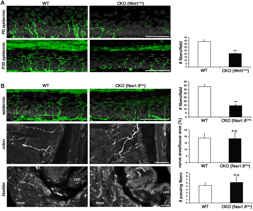
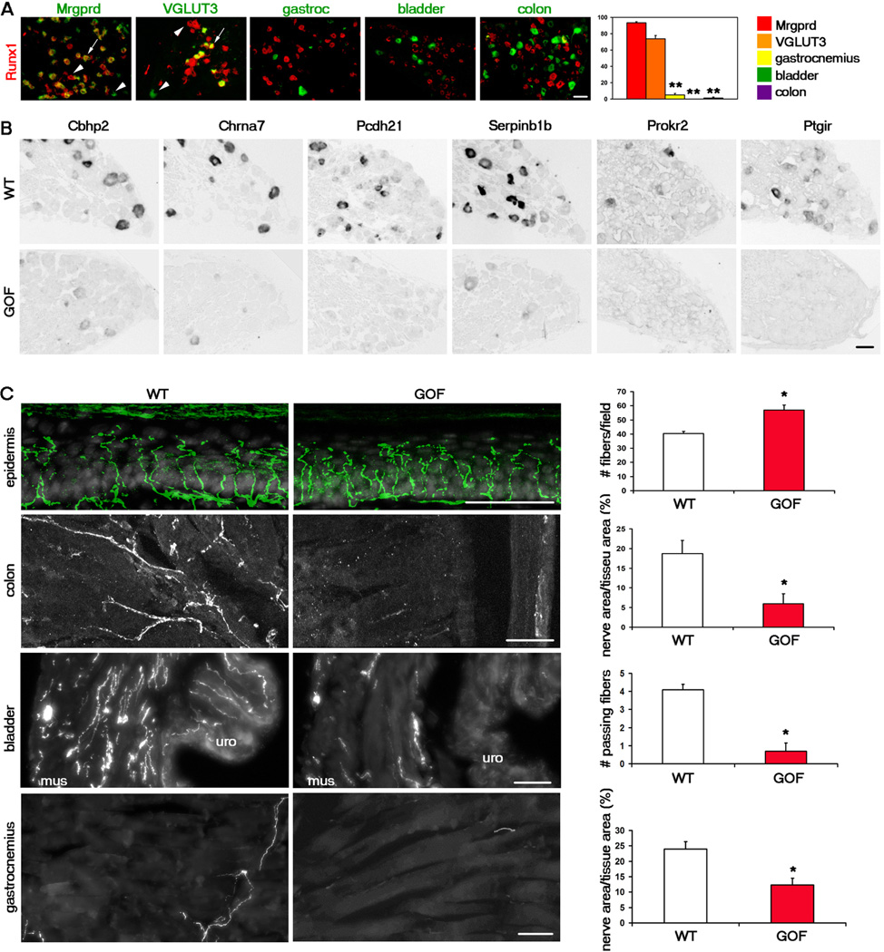
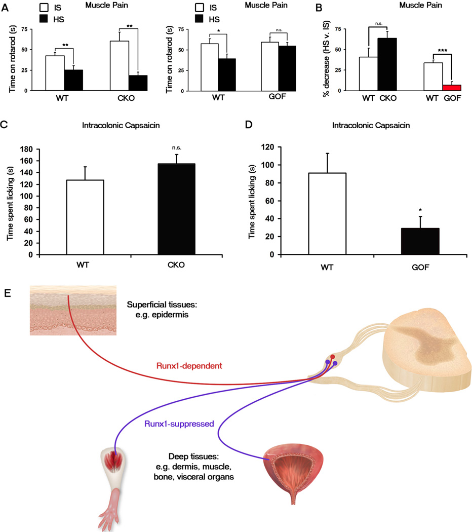
Mammalian pain-related sensory neurons are derived from TrkA lineage neurons located in the dorsal root ganglion. These neurons project to peripheral targets throughout the body, which can be divided into superficial and deep tissues. Here, we find that the transcription factor Runx1 is required for the development of many epidermis-projecting TrkA lineage neurons. Accordingly, knockout of Runx1 leads to the selective loss of sensory innervation to the epidermis, whereas deep tissue innervation and two types of deep tissue pain are unaffected. Within these cutaneous neurons, Runx1 suppresses a large molecular program normally associated with sensory neurons that innervate deep tissues, such as muscle and visceral organs. Ectopic expression of Runx1 in these deep sensory neurons causes a loss of this molecular program and marked deficits in deep tissue pain. Thus, this study provides insight into a genetic program controlling the segregation of cutaneous versus deep tissue pain pathways.


Figures

Figure 1
Figure 1. Cellular characterization of the Runx1-suppressed genes Cbhp2, Chrna7, Pcdh21, Serpinb1b, Prokr2, and Ptgir
(A) In situ hybridization (ISH) with indicated probes on adult lumbar DRG sections of wild-type (“WT”) or Runx1 conditional knockout (CKO) mice. (B) mRNAs of Runx1-suppressed genes (red) detected by ISH were excluded from Mrpgrd+ neurons marked by GFP expression (green, from MrpgrdGFP/+ mice) and from VGLUT3+ neurons marked by Tomato expression (pseudo green, from VGLUT3Cre;RosaTomato fate-mapping mice), but were detected in subsets of neurons retrogradely labeled from the gastrocnemius muscle and/or from the bladder (green). Arrows and arrowheads indicate examples of retrogradely labeled neurons expressing or not expressing the indicated gene, respectively. (C) The percentage of Mrgprd-GFP+, VGLUT3-Tomato+, or neurons retrogradely labeled from the gastrocnemius or bladder that expressed the indicated Runx1-suppressed gene. n ≥ 3 animals for each analysis. Asterisks indicate a significant enrichment of expression in muscle and/or bladder DRG neurons, in comparison with that in Mrgprd-GFP+ or VGLUT3-Tomato+ neurons. Error bars, SEM; * p < 0.05. 924–1124 Mrpgrd-GFP+ cells, 674–1129 VGLUT3-Tomato+ cells, 121–208 muscle retrogradely labeled cells, and 156–364 bladder retrogradely labeled cells were counted for each gene. (D) Genetic marking of the innervation of Prokr2 lineage neurons by using Prokr2Cre;RosaTomato fate-mapping mice (see also Figure S2). Tomato+ nerve terminals of Prokr2 lineage neurons (red) are virtually excluded from the epidermis (“epi”, thin glabrous skin), with cell nuclei revealed by DAPI staining (blue). Note an uncharacterized group of Prokr2-expressing cells in the dermis (“der”). Tomato+ nerve terminals were detected in the gastrocnemius, the bladder, and the colon. For bladder and colon, “mus” for muscular layer, “uro” for urothelial layer, and “muc” for mucosal layer. All scale bars represent 50 µm. See also Figures S1 and S2.
Figure 2
Figure 2. Runx1 is required selectively for sensory innervation to the epidermis
(A) Sensory innervation to the thin glabrous epidermis of the hindpaw at P0 and P30 in the WT (Runx1F/F) and early CKO (Runx1F/F;Wnt1Cre), represented by PGP9.5 staining (green). Keratinocytes are represented by DAPI staining (grey). The number of PGP9.5+ fibers/field of 100 DAPI+ basal keratinocytes at P30 was quantitated. (B) Sensory innervation to indicated regions of WT and late CKO mice at P30, by PGP9.5 staining for epidermis and by GFP immunostaining for colon and bladder. For the epidermis, “WT” is Runx1F/F, and “CKO” is Runx1F/F;Nav1.8Cre. The number of PGP9.5+ fibers/field of 100 DAPI+ basal keratinocytes at P30 was quantitated. For the colon and the bladder, “WT” is Nav1.8Cre;TauLSL-,EGFP/+, and “CKO” is Runx1F/F;Nav1.8Cre;TauLSL-,EGFP/+. The percentages of colon tissue area that is innervated and the number of fibers passing through lines drawn through the muscular layer of the bladder were quantitated. Two methods were used to quantitate bladder innervation, both producing similar results (see Supplementary Methods and Figure S5). n = 3 animals for each genotype. Error bars, SEM; ** p < 0.01; n.s. (not significant) p ≥ 0.05. Methods of quantitation are detailed in Supplementary Experimental Procedures and in Figure S5. All scale bars represent 50 µm.
Figure 3
Figure 3. Downregulation of Runx1 is necessary for the development of deep tissue sensory neurons
(A) Runx1 mRNA (red) was detected in cutaneous neurons marked by Mrgprd-GFP (962 neurons counted) and VGLUT3-Tomato (844 neurons counted), but was largely excluded from neurons retrogradely labeled from the gastrocnemius (129 neurons counted), bladder (329 neurons counted), and colon (338 neurons counted) as indicated by the percentages of these labeled neurons expressing Runx1. Asterisks indicate a significant difference in expression between Mrgprd-GFP+/VGLUT3-Tomato+ neurons and muscle, bladder, or colon neurons. Error bars, SEM; ** p < 0.01. (B) In situ hybridization with indicated probes on adult lumbar DRG sections of wild-type (“WT”, TauLSL-Runx1/+) or Runx1 gain-of-function (“GOF”, Nav1.8Cre;TauLSL-Runx1/+) mice. (C) Sensory innervation to indicated regions of adult WT and GOF mice. For the epidermis, “WT” is TauLSL-Runx1/+, and “GOF” is Nav1.8Cre;TauLSL-Runx1/+. The number of PGP9.5+ fibers/field of 100 DAPI+ basal keratinocytes was quantitated. For the colon, “WT” is Nav1.8Cre;TauLSL-mEGFP/+, and “GOF” is Nav1.8Cre;TauLSL-Runx1/LSL-mEGFP. The percentage of tissue area that is innervated was quantitated. For the bladder and the gastrocnemius, “WT” is Nav1.8Cre;RosaLSL-Tomato/+, and “GOF” is Nav1.8Cre;TauLSL-Runx1/+;RosaLSL-Tomato/+. The number of fibers passing through lines drawn through the muscular layer of the bladder and the percentage of gastrocnemius tissue area that is innervated were quantitated. Two methods were used to quantitate bladder innervation, both producing similar results (see Supplementary Methods and Figure S5). n = 3 animals for each genotype. Error bars, SEM; *p < 0.05. Methods of quantitation are detailed in Supplementary Experimental Procedures and in Figure S5. All scale bars represent 50 µm. See also Figure S3.
Figure 4
Figure 4. Muscle and visceral pain are impaired in Runx1 GOF mice but not in Runx1 CKO mice
(A, B) Muscle pain assay. Time spent on an accelerating rotarod after the injection of hypertonic saline (“HS”) was compared with that after the injection of isotonic saline (“IS”). (A) Runx1 CKO (Runx1F/F;Wnt1Cre) mice (n = 9) displayed a significant decrease in rotatod performance after HS injection as did their WT (Runx1F/F) littermates (n = 9). Runx1 GOF (Nav1.8Cre;TauLSL-Runx1/+) mice (n = 8) displayed no significant decrease after HS injection in contrast to a significant decrease by their WT (TauLSL-Runx1/+) littermates (n = 8). (B) The percentage decrease after HS was calculated for each animal. Shown are the comparisons of the percentage decrease for the CKO and the GOF with their wild-type littermates. Error bars, SEM; * p < 0.05; ** p < 0.01; n.s., p ≥ 0.05. (C, D) Intracolonic capsaicin injection visceral pain assay. (C) No difference in time spent licking between Runx1 CKO (Runx1F/F;Wnt1Cre) mice (n = 7) compared to their WT (Runx1F/F) littermates (n = 6). (D) Runx1 GOF (Nav1.8Cre;TauLSL-Runx1/+) mice (n = 7) displayed a significant decrease in time spent licking compared to their WT (TauLSLRunx1/+) littermates (n = 7). Error bars, SEM; * p < 0.05; n.s., p ≥ 0.05. (E) Dynamic Runx1 expression and activity control the segregation of DRG neurons innervating the superficial ectodermal versus deep mesodermal/endodermal tissues. Neurons expressing Runx1-dependent genes innervate the skin epidermis and hair follicles, tissues fully or partly derived from the ectoderm; these neurons are required to sense cutaneous pain. Neurons expressing Runx1-suppressed genes innervate the mesodermal/endodermal tissues; these neurons are required to sense deep tissue pain. See also Figure S4.

Comment in

Similar articles

Cited by

References

    1. Abdel Samad O, Liu Y, Yang FC, Kramer I, Arber S, Ma Q. Characterization of two Runx1-dependent nociceptor differentiation programs necessary for inflammatory versus neuropathic pain. Mol Pain. 2010;6:45. - PMC - PubMed
    1. Agarwal N, Offermanns S, Kuner R. Conditional gene deletion in primary nociceptive neurons of trigeminal ganglia and dorsal root ganglia. Genesis. 2004;38:122–129. - PubMed
    1. Basbaum AI, Bautista DM, Scherrer G, Julius D. Cellular and molecular mechanisms of pain. Cell. 2009;139:267–284. - PMC - PubMed
    1. Bennett DL, Dmietrieva N, Priestley JV, Clary D, McMahon SB. trkA, CGRP and IB4 expression in retrogradely labelled cutaneous and visceral primary sensory neurones in the rat. Neurosci Lett. 1996;206:33–36. - PubMed
    1. Bove SE, Flatters SJ, Inglis JJ, Mantyh PW. New advances in musculoskeletal pain. Brain Res Rev. 2009;60:187–201. - PMC - PubMed

Publication types

MeSH terms

Substances

LinkOut - more resources