. 2014 Feb:67:426-36.
doi: 10.1016/j.freeradbiomed.2013.11.026. Epub 2013 Dec 4.

Developmental susceptibility of neurons to transient tetrahydrobiopterin insufficiency and antenatal hypoxia-ischemia in fetal rabbits

Affiliations

Developmental susceptibility of neurons to transient tetrahydrobiopterin insufficiency and antenatal hypoxia-ischemia in fetal rabbits

Lei Yu et al. Free Radic Biol Med. 2014 Feb.

Abstract

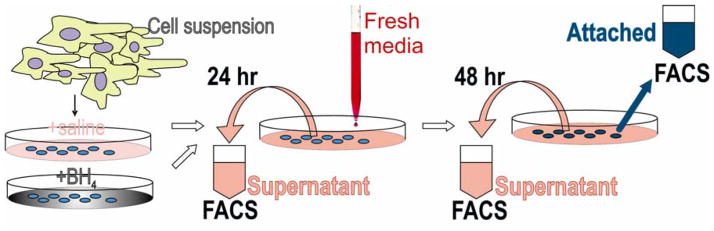
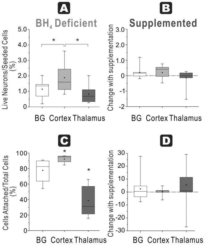
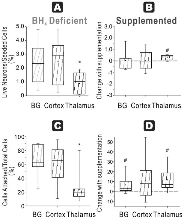
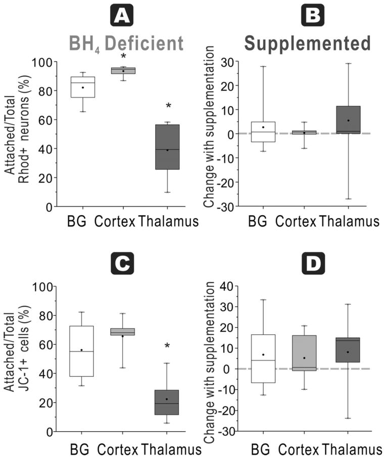
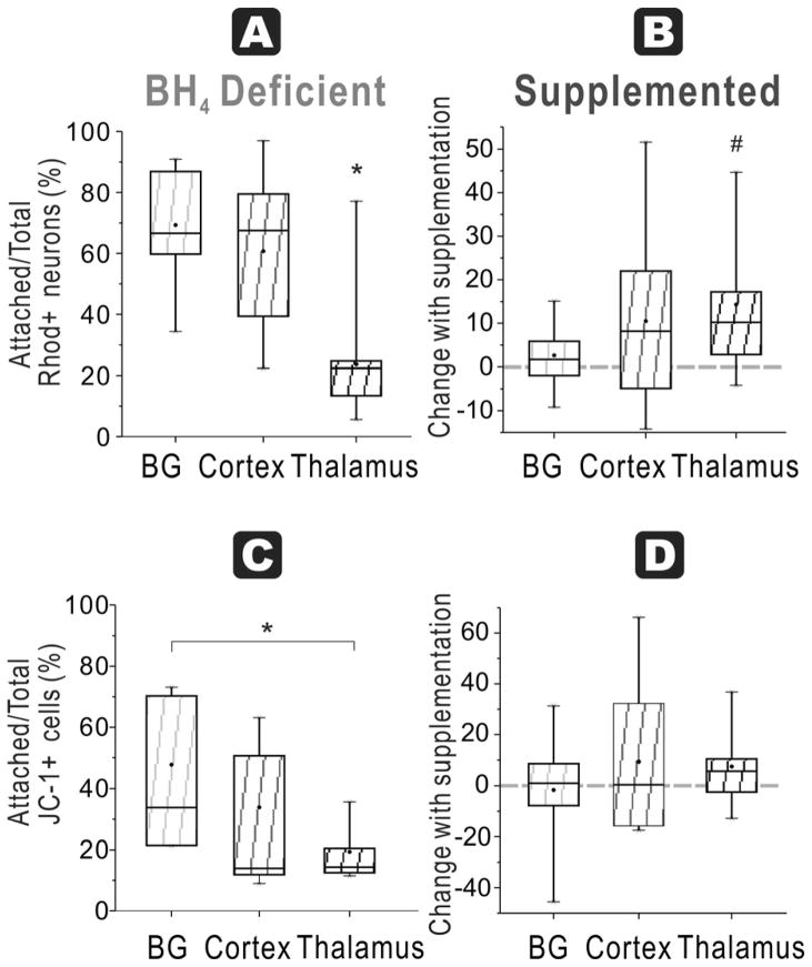
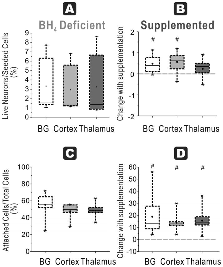
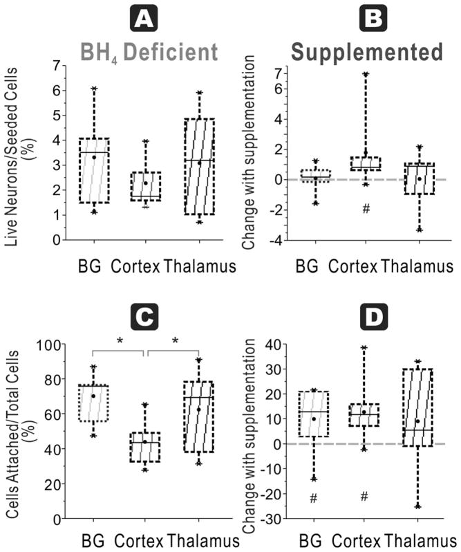
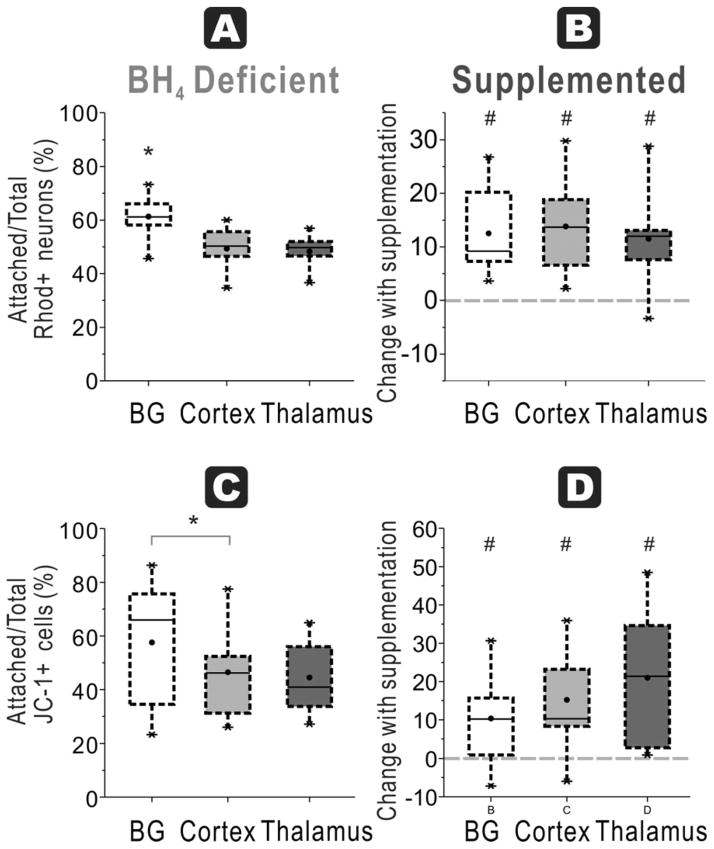
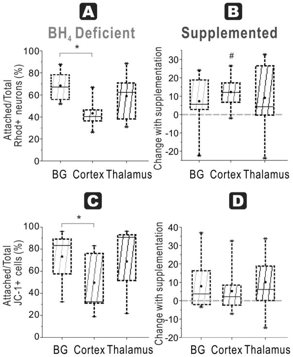
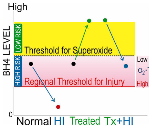
Tetrahydrobiopterin (BH4) is important for normal brain development as congenital BH4 deficiencies manifest movement disorders at various childhood ages. BH4 transitions from very low levels in fetal brains to higher "adult" levels postnatally, with the highest levels in the thalamus. Maternal supplementation with the BH4 precursor sepiapterin reduces postnatal motor deficits and perinatal deaths after 40-min fetal hypoxia-ischemia (HI) at 70% gestation, suggesting that brain BH4 is important in improving function after HI. We tested the hypothesis that the intrinsically low concentrations of BH4 made fetal neurons vulnerable to added insults. Brains were obtained from naïve fetal rabbits or after 40-min HI, at 70% (E22) and 92% gestation (E29). Neuronal cultures were prepared from basal ganglia, cortex, and thalamus, regions with different intrinsic levels of BH4. Cultures were grown with or without added BH4 for 48h. Cell survival and mitochondrial function were determined by flow cytometry. At E22, thalamic cells had the lowest survival rate in a BH4-free milieu, in both control and HI groups, whereas BH4 supplementation ex vivo increased neuronal survival only in HI cells. Neuronal survival was similar in all regions without BH4 at E29. BH4 supplementation increased cell survival and cells with intact mitochondrial membrane potential, from basal ganglia and cortex, but not thalamus. After E29 HI, however, the benefit of BH4 was limited to cortical neurons. We conclude that BH4 is important for fetal neuronal survival after HI especially in the premature thalamus. Supplementation of BH4 has a greater benefit at an earlier gestational age.

Keywords: Anoxia; Basal ganglia; Brain; Cell survival; Cortex; Fetus; Free radicals; Neurons; Premature; Tetrahydrobiopterin; Thalamus.


Conflict of interest statement

Conflict of interest: The authors declare no competing financial interests.

Figures

Figure 1
Figure 1
Experimental Plan and Flow cytometric Analysis Cell suspensions were analyzed and subpopulations of 1) living cells defined by negative propidium iodide staining (PI −), 2) neurons, cells positive for cholera toxin, 3) neurons with functioning mitochondria, cells positive for both cholera toxin and Rhodamine 123 (Cholera + Rhodamine +) as described previously [7], and 4) cells with high mitochondrial function that had higher red than green fluorescence with JC-1 staining. JC-1 was used in a concentration of 5 μg/ml for assessing mitochondrial function [8]. The healthy cells with high mitochondrial function have higher Δψm and JC-1 spontaneously forms complexes known as J-aggregates with intense red fluorescence.
Figure 2
Figure 2. Regional susceptibility to BH4 deficiency for neuronal survival and function at E22
A. Healthy neurons % seeded: In E22 brains, the ratio of live neurons (Cholera toxin and Rhodamine positive cells after 48h culture) divided by total cells (from flow cytometer count of initial cell suspension) show the highest number in cortex compared to basal ganglia and thalamus in a BH4 deficient environment (ANOVA p=0.0153; * p=0.0354 cortex vs basal ganglia, p=0.0028 vs. thalamus, paired t-test). B. With BH4 supplementation, there are no increases in recovery of the ratio (shown as a % change) in any of the groups. C. Attachment efficiency of cells: The efficiency of attachment given by the percentage of attached cells (at 48 h) over the total cells obtained in supernatants at 24 h and 48 hr and attached cells show that the lowest attachment in the thalamus group (ANOVA p<0.0001, *p<0.0001 vs cortex and p=0.007 vs basal ganglia, paired t-test); basal ganglia is lower than cortex (p=0.0116, paired t-test). D. Supplementation with BH4 did not increase the efficiency of attachment in any of the groups (dashed line shows 0 change).
Figure 3
Figure 3. HI at E22 and BH4 dependency on neuronal survival and function
A. Healthy neurons % seeded: In the HI group for E22, ratio of live neurons (Cholera toxin and Rhodamine positive cells after 48h culture) divided by total cells (from flow cytometer count of initial cell suspension) show the lowest number in thalamus in a BH4 deficient environment (ANOVA p=0.0172; *p =0.0009 vs basal ganglia and p=0.0012 vs cortex, paired t-test). B. Healthy neurons % seeded With BH4 supplementation, this ratio was significantly increased in thalamus, but not in basal ganglia and cortex groups (# p=0.0004, paired t-test). C. Attachment efficiency of cells: In the HI group, the efficiency of attachment given by the percentage of attached cells (at 48 h) over the total cells obtained in supernatants at 24 h and 48 hr and attached cells show that the lowest attachment in the thalamus group (ANOVA p=0.0003; * p=0.0004 vs. basal ganglia and p=0.0021 vs cortex, n=11, paired t-test). D. Attachment efficiency of cells: Supplementation with BH4 increase the efficiency of attachment in both basal ganglia (# p=0.0398) and thalamus (# p=0.0056, n=11, paired t-tests).
Figure 4
Figure 4. BH4-dependent mitochondrial function and regional susceptibility at E22
A. Attachment efficiency of functioning mitochondria neurons: In E22 brains, the ratio of attached neurons with functioning mitochondria (Rhodamine + Cholera + cells at 48 h) over total neurons with functioning mitochondria (in supernatant at 24 and 48 h plus attached at 48 h) was the highest in the cortex in a BH4 deficient environment (ANOVA p<0.0001; *p=0.0057 cortex vs basal ganglia, p<0.0001 cortex vs. thalamus, p=0.0003 basal ganglia vs. thalamus, paired t-test). B. With BH4 supplementation, there are no increases in recovery of the ratio (shown as a % change) in any of the groups. C. Attachment efficiency of high mitochondria cells: Thalamus neurons from E22 rabbit fetal brain had the lowest ratio of attached cells high mitochondrial function (JC healthy -1 red>green fluorescent cells at 48 h) to total cells with high mitochondrial function (in supernatant at 24 and 48 h plus attached at 48 h) without BH4 supplementation (ANOVA p<0.0001; * p=0.0068 vs. basal ganglia and p=0.0001 vs. cortex, paired t-test). D. There were no differences in this ratio in any of the groups with BH4 supplementation.
Figure 5
Figure 5. HI at E22 and BH4-dependent regional mitochondrial function
A. In the HI group for E22, the ratio of attached neurons with functioning mitochondria (Rhodamine + Cholera + cells at 48 h) over total neurons with functioning mitochondria (in supernatant at 24 and 48 h plus attached at 48 h) was the lowest in thalamus in a BH4 deficient environment (ANOVA p<0.0001; * p=0.0002 vs basal ganglia and p=0.0007 vs cortex, paired t-test). B. Attachment efficiency of functioning mitochondria neurons: With BH4 supplementation, this ratio significantly increased in thalamus (# p=0.0099, paired t-test) but not in basal ganglia and cortex. C. Attachment efficiency of high mitochondria cells: In the E22 HI group, thalamus had the lowest ratio of attached cells with healthy mitochondria (JC-1 red>green fluorescent cells at 48 h) to total cells with healthy mitochondria (in supernatant at 24 and 48 h plus attached at 48 h) without BH4 supplementation (ANOVA p=0.0313; *p=0.0098 thalamus vs basal ganglia, paired t-test). D. With BH4 supplementation, there are no increases in recovery of this ratio in any of the groups.
Figure 6
Figure 6. Most regions show improvement with BH4 in neuronal survival and function at E29
A. Healthy neurons % seeded: In E29 brains (depicted by dashed lines), the ratio of live neurons (Cholera toxin and Rhodamine positive cells after 48h culture) divided by total cells (from flow cytometer count of initial cell suspension) was not different among cortex, basal ganglia and thalamus in a BH4 deficient environment. B. Healthy neurons % seeded: With BH4 supplementation, this ratio significantly increased in both basal ganglia and cortex (# p=0.0109 and p=0.0103 respectively, paired t-test) but not in thalamus group. C. Attachment efficiency of cells: The efficiency of attachment given by the percentage of attached cells (at 48 hr) over the total cells obtained in supernatants at 24 h and 48 hr and attached cells at 48 hr showed no difference attachment among the thalamus, cortex and basal ganglia. D. Attachment efficiency of cells: Supplementation with BH4 increased the efficiency of attachment in all of the groups (#BG p=0.0084, Cortex p=0.0006, thalamus p=0.0012, paired t-test).
Figure 7
Figure 7. HI at E29 and BH4 dependency on neuronal survival and function specific to cortex
A. Healthy neurons % seeded: In the HI group for E29, ratio of live neurons (Cholera toxin and Rhodamine positive cells after 48h culture) divided by total cells (from flow cytometer count of initial cell suspension showed no difference among the three regions. B. Healthy neurons % seeded: With BH4 supplementation, this ratio was significantly increased in cortex (#, p=0.04911, n=9/group, paired t-test), but not in basal ganglia and thalamus. C. Attachment efficiency of cells: In the HI group, the efficiency of attachment given by the percentage of attached cells (at 48 h) over the total cells obtained in supernatants at 24 h and 48 hr and attached cells showed that cortex had the least cells (ANOVA p=0.0132; * p=0.0015 cortex vs. basal ganglia, p=0.0399 cortex vs. thalamus, n=9/group, paired t-test). D. Attachment efficiency of cells: Supplementation with BH4 increased the efficiency of attachment in both basal ganglia and cortex (# p =0.0421 and p=0.0126 respectively, n=9, paired t-test), but not in thalamus.
Figure 8
Figure 8. BH4-dependent mitochondrial function in all regions at E29
A. In E29 brains, the ratio of attached neurons with functioning mitochondria (Rhodamine + Cholera + cells at 48 h) over total neurons with functioning mitochondria (in supernatant at 24 and 48 h plus attached at 48 h) show the highest number in basal ganglia compared to cortex and thalamus in a BH4 deficient environment (ANOVA p=0.0030; *p=0.0017 basal ganglia vs. cortex, p=0.0072 vs. thalamus, n=9/group, paired t-test). B. Attachment efficiency of functioning mitochondria neurons: With BH4 supplementation, there are significant increases in recovery of the ratio (shown as a % change) in all groups (# basal ganglia p=0.0017, Cortex p=0.0013, thalamus p=0.0045, n=9/group, paired t-test). C. Attachment efficiency of high mitochondria cells: Basal ganglia neurons from E29 rabbit fetal brain had higher ratio of attached cells with high mitochondrial function (JC-1 red>green fluorescent cells at 48 h) to total cells with high mitochondrial function (in supernatant at 24 and 48 h plus attached at 48 h) compared to cortex, without BH4 supplementation (* p=0.0234, n=9/group, paired t-test but ANOVA not significant). D. Attachment efficiency of high mitochondria cells: With BH4 supplementation, there were significant increases in recovery of this ratio in all the groups (# basal ganglia p=0.0314, Cortex p=0.0102, thalamus p=0.0079, n=9/group, paired t-test).
Figure 9
Figure 9. HI at E29 and BH4-dependent mitochondrial function specific to cortex
A. Attachment efficiency of functioning mitochondria neurons: In the HI group for E29, the ratio of attached neurons with functioning mitochondria (Rhodamine + Cholera + cells at 48 h) over total neurons with functioning mitochondria (in supernatant at 24 and 48 h plus attached at 48 h) in basal ganglia was higher than cortex in a BH4 deficient environment (ANOVA p=0.0137; * p=0.0077 basal ganglia vs. cortex, paired t-test). B. Attachment efficiency of functioning mitochondria neurons: With BH4 supplementation, this ratio significantly increased in cortex (# p=0.0046, paired t-test) but not in basal ganglia and thalamus. C. Attachment efficiency of high mitochondria cells: Cortex had a lower ratio of attached cells with healthy mitochondria (JC-1 red>green fluorescent cells at 48 h) to total cells with healthy mitochondria (in supernatant at 24 and 48 h plus attached at 48 h) compared to basal ganglia without BH4 supplementation (* p=0.0179, paired t-test although ANOVA not significant). D. With BH4 supplementation, there were no increases in recovery of this ratio in any of the groups.
Figure 10
Figure 10. Double-hit hypothesis as proposed by Vasquez-Vivar and Tan
BH4=Tetrahydrobiopterin, O2·− =superoxide, Tx=treated, HI=hypoxia-ischemia. Note that even in high risk phase another insult other than HI can result in injury.

References

    1. Ichinose H, Nomura T, Sumi-Ichinose C. Metabolism of tetrahydrobiopterin: its relevance in monoaminergic neurons and neurological disorders. Chem Rec. 2008;8:378–85. - PubMed
    1. Fink JK, Ravin P, Argoff CE, Levine RA, Brady RO, Hallett M, et al. Tetrahydrobiopterin administration in biopterin-deficient progressive dystonia with diurnal variation. Neurology. 1989;39:1393–5. - PubMed
    1. Frye RE, DeLatorre R, Taylor HB, Slattery J, Melnyk S, Chowdhury N, et al. Metabolic effects of sapropterin treatment in autism spectrum disorder: a preliminary study. Transl Psychiatry. 2013;3:e237. - PMC - PubMed
    1. Danfors T, von Knorring AL, Hartvig P, Langstrom B, Moulder R, Stromberg B, et al. Tetrahydrobiopterin in the treatment of children with autistic disorder: a double-blind placebo-controlled crossover study. J Clin Psychopharmacol. 2005;25:485–9. - PubMed
    1. Vasquez-Vivar J, Whitsett J, Derrick M, Ji X, Yu L, Tan S. Tetrahydrobiopterin in the prevention of hypertonia in hypoxic fetal brain. Ann Neurol. 2009;66:323–31. - PMC - PubMed

Publication types

MeSH terms