. 2014 Feb:59:229-34.
doi: 10.1016/j.bone.2013.11.026. Epub 2013 Dec 4.

Ovariectomy enhances mechanical load-induced solute transport around osteocytes in rat cancellous bone

Affiliations

Ovariectomy enhances mechanical load-induced solute transport around osteocytes in rat cancellous bone

Cesare Ciani et al. Bone. 2014 Feb.

Abstract

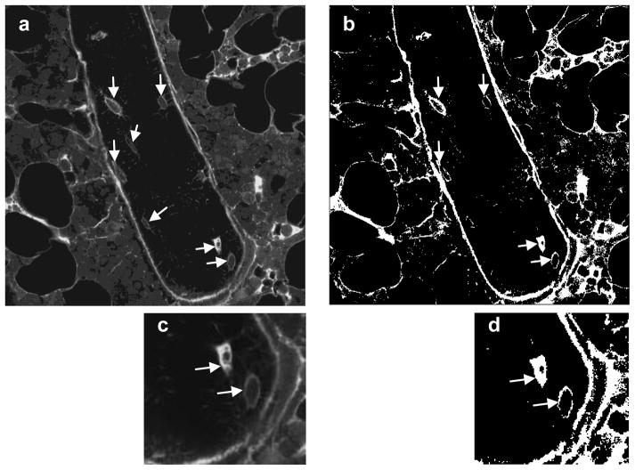
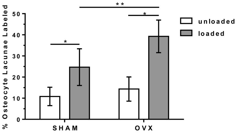
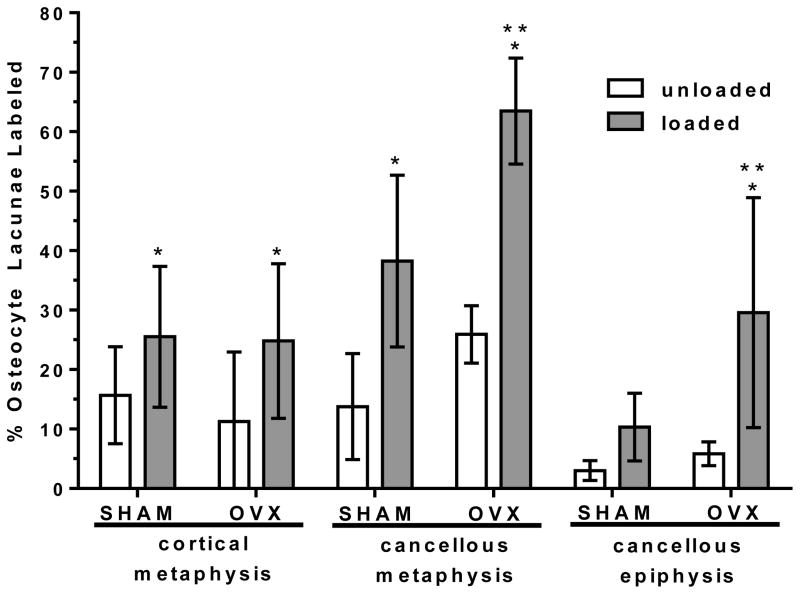
To test if osteoporosis alters mechanical load-induced interstitial fluid flow in bone, this study examined the combined effect of estrogen deficiency and external loading on solute transport around osteocytes. An in vivo tracer, FITC-labeled bovine serum albumin, was injected into anesthetized ovariectomized and control female Sprague-Dawley rats before the right tibia was subjected to a controlled, physiological, non-invasive sinusoidal load to mimic walking. Tracer movement through the lacunar-canalicular system surrounding osteocytes was quantified in cortical and cancellous bone from the proximal tibia using confocal microscopy, with the non-loaded tibia serving as internal control. Overall, the application of mechanical loading increased the percentage of osteocyte lacunae labeled with injected tracer, and ovariectomy further enhanced movement of tracer. An analysis of separate regions demonstrated that ovariectomy enhanced in vivo transport of the injected tracer in the cancellous bone of the tibial epiphysis and metaphysis but not in the cortical bone of the metaphysis. These findings show that bone changes due to reduced estrogen levels alter convectional transport around osteocytes in cancellous bone and demonstrate a functional difference of interstitial fluid flow around osteocytes in estrogen-deficient rats undergoing the same physical activity as controls. The altered interstitial fluid flow around osteocytes is likely related to nanostructural matrix-mineral level differences recently demonstrated at the lacunar-canalicular surface of estrogen-deficient rats, which could affect the transmission of mechanical loads to the osteocyte.

Keywords: Canaliculi; Interstitial fluid; Mechanotransduction; Osteocyte; Osteoporosis; Perilacunar remodeling.


Conflict of interest statement

Conflict of interest statement

The authors have no conflicts of interest.

Figures

Figure 1
Figure 1
(a) Sinusoidal loading was applied non-invasively to the rat lower hindlimb, which was constrained between a custom-made knee cup and foot holder. The lower limb was compressed in the direction indicated by the arrows. (b) Strain-gage calibration of the loading device using control rats (n=3; each rat represented by a symbol, ● ▲ ◆) illustrates the relationship between applied load (N) and measured deformation (microstrain) of the proximal tibia. After tracer injection, an applied load of 14 N, which engenders ~500 microstrain at the medial proximal diaphysis, was applied at 1 Hz to mimic slow walking.
Figure 2
Figure 2
Scanning electron microscopy image of a rat proximal tibia frontal section illustrating where the cortical and cancellous bone confocal images were taken to assess osteocyte lacunae labeled with injected tracer. For the metaphyseal region, five cancellous bone image samples (187.5 μm × 187.5 μm images, smaller boxes) were taken in the region spanning 1 to 3 mm distal to the growth plate; primary spongiosa were not included. Ten cortical metaphyseal image samples (375 μm × 375 μm images, larger boxes) were taken from the cortex region. For the epiphyseal region, three image samples were taken right above the growth plate, without including any region of the growth plate, to ensure consistency in sampling, and two additional regions of interest were sampled more proximally (187.5 μm × 187.5 μm images, smaller boxes).
Figure 3
Figure 3
Tracer transport through the lacunar-canalicular system was assessed using confocal microscopy to identify osteocyte lacunae considered labeled in both loaded and unloaded tibiae. (a) A typical grayscale image of cancellous bone of the metaphysis. The trabecula shown is surrounded by bone marrow, and the fully visible osteocytes are indicated with arrows. (b) The same image after thresholding; labeled osteocyte lacunae are marked with arrows. (c) Higher magnification of the image in (a) showing two osteocytes, and (d) the same image after thresholding. The arrows in (d) indicate that both osteocytes satisfied the criteria to be counted as labeled (osteocyte lacunae presenting at least 80% of the lacunar space stained or 80% of the periphery of the lacuna stained).
Figure 4
Figure 4
The percentage of osteocyte lacunae labeled with injected tracer (mean ± SD) for the SHAM and OVX groups with the measurements at three anatomical locations (cortical metaphysis, cancellous metaphysis, and cancellous epiphysis) pooled for each animal for the loaded and contralateral, unloaded tibiae. * significant difference compared to unloaded in same group (p < 0.05); ** significant difference compared to loaded SHAM (p < 0.05).
Figure 5
Figure 5
The percentage of osteocyte lacunae labeled with injected tracer (mean ± SD) for the SHAM and OVX groups for the three anatomical locations analyzed: cortical metaphysis, cancellous metaphysis, and cancellous epiphysis in the loaded and contralateral, unloaded tibiae. * significant difference compared to unloaded in same group (p < 0.05); ** significant difference compared to loaded SHAM at same location (p < 0.05).

References

    1. Hancox NM. Biology of Bone. Cambridge, England: Cambridge University Press; 1972.
    1. Cowin SC, Moss-Salentijn L, Moss ML. Candidates for the mechanosensory system in bone. J Biomech Eng. 1991;113:191–7. - PubMed
    1. Lanyon LE. Osteocytes, strain detection, bone modeling and remodeling. Calcif Tissue Int. 1993;53 (Suppl 1):S102–6. - PubMed
    1. Qin YX, Lin W, Rubin C. The pathway of bone fluid flow as defined by in vivo intramedullary pressure and streaming potential measurements. Ann Biomed Eng. 2002;30:693–702. - PubMed
    1. Fritton SP, Weinbaum S. Fluid and solute transport in bone: flow-induced mechanotransduction. Annu Rev Fluid Mech. 2009;41:347–74. - PMC - PubMed

Publication types

LinkOut - more resources