. 2014 Mar:140:56-62.
doi: 10.1016/j.jsbmb.2013.11.017. Epub 2013 Dec 5.

Activation of vitamin D receptor promotes VEGF and CuZn-SOD expression in endothelial cells

Affiliations

Activation of vitamin D receptor promotes VEGF and CuZn-SOD expression in endothelial cells

Weijie Zhong et al. J Steroid Biochem Mol Biol. 2014 Mar.

Abstract

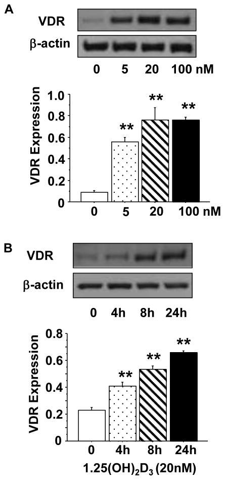
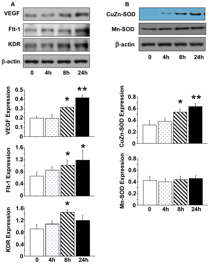
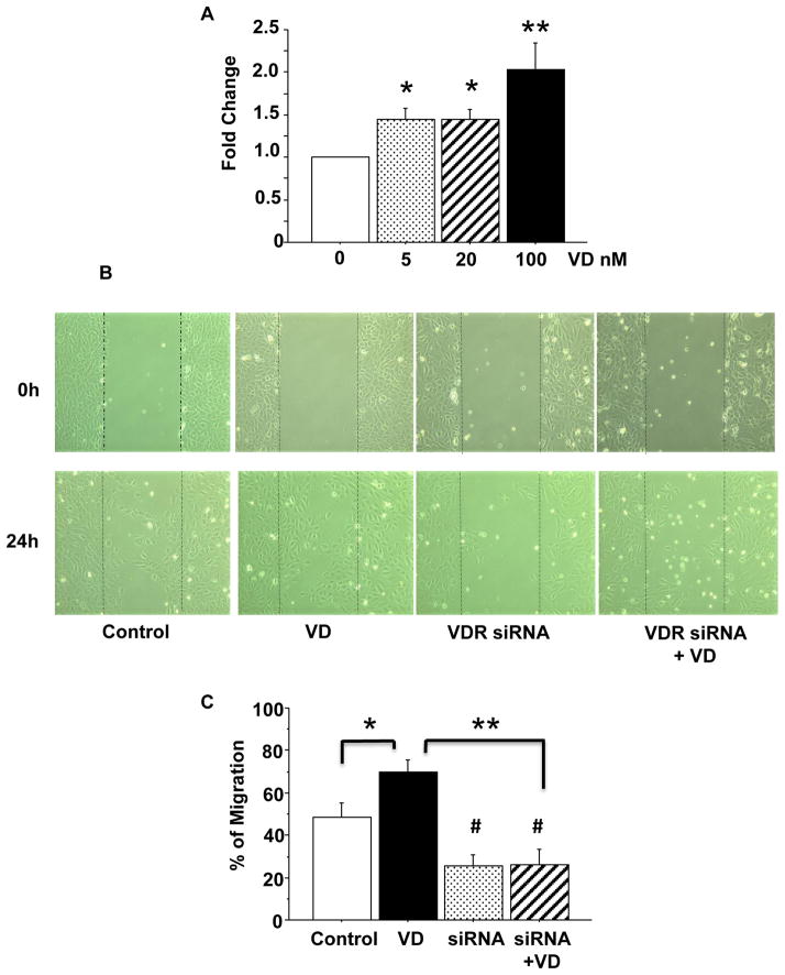
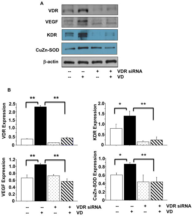
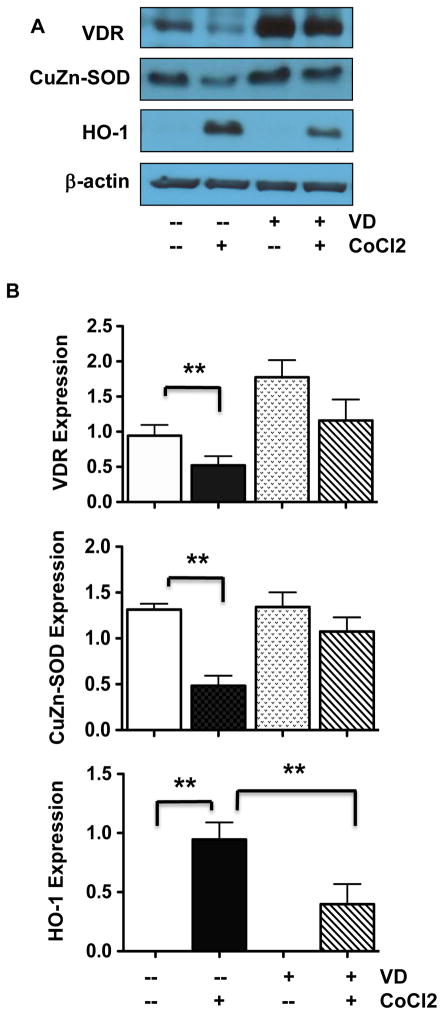
Endothelial dysfunction associated with vitamin D deficiency has been linked to many chronic vascular diseases. Vitamin D elicits its bioactive actions by binding to its receptor, vitamin D receptor (VDR), on target cells and organs. In the present study, we investigated the role of VDR in response to 1,25(OH)₂D₃ stimulation and oxidative stress challenge in endothelial cells. We found that 1,25(OH)₂D₃ not only induced a dose- and time-dependent increase in VDR expression, but also induced up-regulation of vascular endothelial growth factor (VEGF) and its receptors (Flt-1 and KDR), as well as antioxidant CuZn-superoxide dismutase (CuZn-SOD) expression in endothelial cells. We demonstrated that inhibition of VDR by VDR siRNA blocked 1,25(OH)₂D₃ induced increased VEGF and KDR expression and prevented 1,25(OH)₂D₃ induced endothelial proliferation/migration. Using CoCl₂, a hypoxic mimicking agent, we found that hypoxia/oxidative stress not only reduced CuZn-SOD expression, but also down-regulated VDR expression in endothelial cells, which could be prevented by addition of 1,25(OH)₂D3 in culture. These findings are important indicating that VDR expression is inducible in endothelial cells and oxidative stress down-regulates VDR expression in endothelial cells. We conclude that sufficient vitamin D levels and proper VDR expression are fundamental for angiogenic and oxidative defense function in endothelial cells.

Keywords: Angiogenic property; CuZn-SOD; Endothelial cells; Oxidative stress; VDR.


Figures

Figure 1
Figure 1
1,25(OH)2D3 induced up-regulation of VDR protein expression in endothelial cells. A: Cells were treated with 1,25(OH)2D3 at concentrations of 0, 5, 20, and 100nM for 24 hours. 1,25(OH)2D3 induced a dose-dependent increase in VDR expression. B: Cells were treated with 20nM of 1,25(OH)2D3 for 0, 4, 8, and 24 hours. 1,25(OH)2D3 induced a time-dependent increase in VDR expression. The bar graphs show the mean ± SE of relative VDR expression after normalized with β-actin expression in each sample, **p<0.01: treated vs. controls. Data are expressed as the mean ± SE from 3 independent experiments.
Figure 2
Figure 2
Effects of 1,25(OH)2D3 on protein expression of VEGF, Flt-1, KDR, CuZn-SOD, and Mn-SOD in endothelial cells. A: Protein expression of VEGF, Flt-1, and KDR in endothelial cells treated with 20nM of 1,25(OH)2D3 for 0, 4, 8, and 24 hours. B: Protein expression of CuZn-SOD and Mn-SOD in endothelial cells treated with 20nM of 1,25(OH)2D3 for 0, 4, 8, and 24 hours. The bar graphs show relative target protein expression after normalized with β-actin expression in each sample, *p<0.05 and **p<0.01 in cells treated with 1,25(OH)2D3 vs. not. Data are expressed as mean ± SE from 3 independent experiments.
Figure 3
Figure 3
1,25(OH)2D3 induced cell proliferation and migration. A: 1,25(OH)2D3 induced endothelial proliferation was determined by MTT assay. Cells were treated with 1,25(OH)2D3 (VD) at concentrations of 0, 5, 20, 100nM for 24 hours (n=6). 1,25(OH)2D3 induced increased cell proliferation was dose-dependent, *p<0.05, and **p<0.01. B: Representative images of 1,25(OH)2D3 induced cell migration (wound healing assay). Images were taken immediately after scratching (0 hour) and 24 hours after addition of 1,25(OH)2D3 in culture. C: Bar graph shows quantitative measure of cell migration. Data are expressed as mean ± SE of wound healing assay from 3 independent experiments. VD: 1,25(OH)2D3; *p<0.05: VD treated vs. untreated control; # p<0.05: VDR siRNA vs. untreated control; and **p<0.01: VDR siRNA or VDR siRNA + VD vs. VD, respectively.
Figure 4
Figure 4
Effects of VDR inhibition on VEGF, KDR, and CuZn-SOD protein expression. VDR siRNA was used to inhibit VDR expression. VDR siRNA not only inhibited 1,25(OH)2D3 induced increased VDR expression, but also blocks 1,25(OH)2D3 induced VEGF, KDR, and CuZn-SOD up-regulation, in endothelial cells. A: VDR, VEGF, KDR, and CuZn-SOD expression in control cells, cells treated with 1,25(OH)2D3, and cells transfected with VDR siRNA with or without addition of 1,25(OH)2D3. B: Relative VDR, VEGF, KDR, and CuZn-SOD expression normalized with β-actin expression, *p<0.05 and **p<0.01. These results indicate that VDR modulates VEGF, KDR, and CuZn-SOD expression in endothelial cells. Data are expressed as the mean ± SE from 3 independent experiments. VD: 1,25(OH)2D3.
Figure 5
Figure 5
Effects of oxidative stress on VDR, CuZn-SOD, and HO-1 protein expression. A: Representative blots for VDR, CuZn-SOD, and HO-1 expression in cells treated with CoCl2 in the presence or absence of 1,25(OH)2D3 in culture. B: Relative protein expression for VDR, CuZn-SOD, and HO-1 after normalized with β-actin expression in each sample, *p<0.05 and **p<0.01. Data are expressed as the mean ± SE from 3 independent experiments.

References

    1. Brumbaugh PF, Haussler MR. Specific binding of 1α,25-dihydroxycholecalciferol to nuclear components of chick intestine. J Biol Chem. 1975;250:1588–1594. - PubMed
    1. McDonnell DP, Mangelsdorf DJ, Pike JW, Haussler MR, O’Malley BW. Molecular cloning of complementary DNA encoding the avian receptor for vitamin D. Science. 1987;235:1214–1217. - PubMed
    1. Bouillon R, Carmeliet G, Verlinden L, van Etten E, Verstuyf A, Luderer HF, Lieben L, Mathieu C, Demay M. Vitamin D and human health: lessons from vitamin D receptor null mice. Endocr Rev. 2008;29:726–776. - PMC - PubMed
    1. Motiwala SR, Wang TJ. Vitamin D and cardiovascular disease. Curr Opin Nephrol Hypertens. 2011;20:345–353. - PubMed
    1. Awad AB, Alappat L, Valerio M. Vitamin D and metabolic syndrome risk factors: evidence and mechanisms. Crit Rev Food Sci Nutr. 2012;52:103–112. - PubMed

Publication types

MeSH terms

LinkOut - more resources