. 2014 Feb:252:105-13.
doi: 10.1016/j.expneurol.2013.11.023. Epub 2013 Dec 6.

A tetra(ethylene glycol) derivative of benzothiazole aniline ameliorates dendritic spine density and cognitive function in a mouse model of Alzheimer's disease

Affiliations

A tetra(ethylene glycol) derivative of benzothiazole aniline ameliorates dendritic spine density and cognitive function in a mouse model of Alzheimer's disease

Jung Min Song et al. Exp Neurol. 2014 Feb.

Abstract

We recently reported that the tetra(ethylene glycol) derivative of benzothiazole aniline, BTA-EG4, acts as an amyloid-binding small molecule that promotes dendritic spine density and cognitive function in wild-type mice. This raised the possibility that BTA-EG4 may benefit the functional decline seen in Alzheimer's disease (AD). In the present study, we directly tested whether BTA-EG4 improves dendritic spine density and cognitive function in a well-established mouse model of AD carrying mutations in APP, PS1 and tau (APPswe;PS1M146V;tauP301L, 3xTg AD mice). We found that daily injections of BTA-EG4 for 2 weeks improved dendritic spine density and cognitive function of 3xTg AD mice in an age-dependent manner. Specifically, BTA-EG4 promoted both dendritic spine density and morphology alterations in cortical layers II/III and in the hippocampus at 6-10 months of age compared to vehicle-injected mice. However, at 13-16 months of age, only cortical spine density was improved without changes in spine morphology. The changes in dendritic spine density correlated with Ras activity, such that 6-10 month old BTA-EG4 injected 3xTg AD mice had increased Ras activity in the cortex and hippocampus, while 13-16 month old mice only trended toward an increase in Ras activity in the cortex. Finally, BTA-EG4 injected 3xTg AD mice at 6-10 months of age showed improved learning and memory; however, only minimal improvement was observed at 13-16 months of age. This behavioral improvement corresponds to a decrease in soluble Aβ 40 levels. Taken together, these findings suggest that BTA-EG4 may be beneficial in ameliorating the synaptic loss seen in early AD.

Keywords: 3xTg AD mice; AD; Alzheimer's disease; Aβ; BTA-EG(4); Dendritic spine; Ras signaling; amyloid-β.


Figures

Fig. 1
Fig. 1
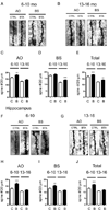
BTA-EG4 increases dendritic spine density in 3xTg AD mice. (A–E) Representative Golgi-stained dendritic segments of cortical layer II/III pyramidal neurons from 6–10 months of age (A) or 13–16 months of age (B) 3xTg AD mice treated with BTA-EG4 (“B”) or vehicle (“C”) control. Quantification of averaged spine densities on apical oblique (AO) (C), basal (BS) (D), and total (AO+BS) (E) dendrites (n=5 brains/group; **p<0.01, ***p<0.001). (F–J) Representative Golgi-stained dendritic segments of cortical layer II/III pyramidal neurons from 6–10 months of age (F) or 13–16 months of age (G) 3xTg AD mice treated with BTA-EG4 or vehicle control. Quantification of averaged spine densities on apical oblique (AO, H), basal (BS, I), and total (AO+BS, J) dendrites (n=3 brains/group; **p<0.01, ***p<0.001).
Fig. 2
Fig. 2
BTA-EG4 alters dendritic spine morphology in 6–10 month-old, but not 13–16 month old, 3xTg AD mice. (A–D) Dendritic spine morphology as a cumulative distribution plot of spine head width (A,C) and spine length (B,D) in cortical layers II/III (A–B) and hippocampal region CA1 (C–D) in 6–10 month old mice treated with BTA-EG4 (Kolmogorov-Smirnov test, n=5 brains/group; *p<0.05). (E–H) Dendritic spine morphology as a cumulative distribution plot of spine head width (E,G) and spine length (F,H) in cortical layers II/III (E–F) and hippocampal region CA1 (G–H) at 13–16 month old mice treated with BTA-EG4 (Kolmogorov-Smirnov test, n=3 brains/group; *p<0.05). (I–L) Summary of the average width of dendritic spines in the cortex (I) and hippocampal CA1 region (K) following BTA-EG4 treatment as a percentage of control levels for each age. Summary of the averaged dendritic spine lengths in the cortex (J) and hippocampal CA3 (L) following BTA-EG4 (“B”) or control (“C”) treatment for 2 weeks a percentage of control levels for each age. ***p < 0.001.
Fig. 3
Fig. 3
BTA-EG4 increases Ras activity and RasGRF1 levels in 6–10 month old 3xTg AD mice. GST-Raf1-RBD pull-down of active Ras from brain lysates of cortex and hippocampus from 6–10 month old (A–D) and 13–16 month old (E-H) 3xTg AD mice injected with control or BTA-EG4 (n=3 brains/group); *p<0.05. (I–L) Western blot of RasGRF1 in brain lysates from cortex (I, J) and hippocampus (K, L) from 6–10 month old 3xTg AD mice i.p. injected with vehicle control (“C”) or BTA-EG4 (“B”) (n=4 brains/group). (M–Q) Western blot of RasGRF1 from cortex (M, O) and hippocampus (P, Q) from 13–16 month old 3xTg AD mice injected with BTA-EG4 or vehicle (n=4 brains/group). β-actin is used as a loading control; *p<0.05.
Fig. 4
Fig. 4
BTA-EG4 injected mice had increased AMPA receptor subunit GluA2 expression at 6–10 months of age. (A–D) Western blot of GluA1 and GluA2 levels in brain lysates from cortex (A, B) and hippocampus (C, D) of 6–10 month old 3x Tg AD mice injected with BTA-EG4 (“B”) or control vehicle solution (“C”) daily for 2 weeks (n=4 brains/group). (E–H) Western blot of levels of GluA1 and GluA2 in brain lysates from cortex (E, F) and hippocampus (G,H) of 13–16 month old 3x Tg AD mice i.p. injected with BTA-EG4 (“B”) or control (“C”) (n=4 brains/group).
Fig. 5
Fig. 5
BTA-EG4 improves cognitive performance of 3xTg AD mice. Spatial learning was tested by Morris water maze in 3xTg mice aged 2–3 months (n = 7 / group), 6–10 months (n = 10 / group), and 13–16 months (n = 7 / group). Escape latencies during the 4-day training phase (A, E, I), swim speed velocity during the training trials (B, F, J), percent time spent in the target quadrant measured during the probe test on day 5 (C, G, K), and the number of platform crossings during probe trial on day 5 (D, H, L) were compared between vehicle (CTRL) and BTA-EG4 (BTA) treated 2–3 month old (A–D), 6–10 month old (E–H), and 13–16 month old (I–L) 3xTg AD mice (*p<0.05, **p<0.01). Aβ ELISA was conducted to compare Aβ levels in 2–3 month old (M, n=7/group), 6–10 month old (N, n=10/group), and 13–16 month old (O, n=4–5/group) 3xTg AD mice injected with BTA-EG4 or vehicle daily for 2 weeks.

References

    1. Capule CC, Yang J. Enzyme-Linked Immunosorbent Assay-Based Method to Quantify the Association of Small Molecules with Aggregated Amyloid Peptides. Analytical Chemistry. 2012;84:1786–1791. - PMC - PubMed
    1. Finder VH, Glockshuber R. Amyloid-beta aggregation. Neurodegener Dis. 2007;4:13–27. - PubMed
    1. Gu Y, Stornetta RL. Synaptic plasticity, AMPA-R trafficking, and Ras-MAPK signaling. Acta Pharmacol Sin. 2007;28:928–936. - PubMed
    1. Habib LK, Lee MT, Yang J. Inhibitors of catalase-amyloid interactions protect cells from beta-amyloid-induced oxidative stress and toxicity. J Biol Chem. 2010;285:38933–38943. - PMC - PubMed
    1. Hsieh H, Boehm J, Sato C, Iwatsubo T, Tomita T, Sisodia S, Malinow R. AMPAR removal underlies Abeta-induced synaptic depression and dendritic spine loss. Neuron. 2006;52:831–843. - PMC - PubMed

Publication types

MeSH terms