. 2014 Feb;10(2):149-55.
doi: 10.1038/nchembio.1408. Epub 2013 Dec 8.

Protonation drives the conformational switch in the multidrug transporter LmrP

Affiliations

Protonation drives the conformational switch in the multidrug transporter LmrP

Matthieu Masureel et al. Nat Chem Biol. 2014 Feb.

Abstract

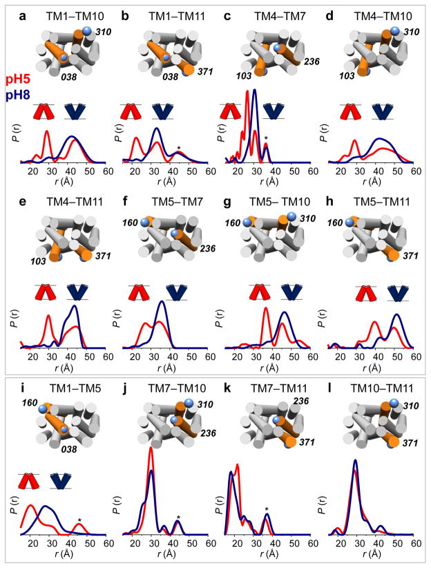
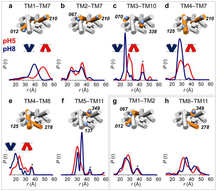
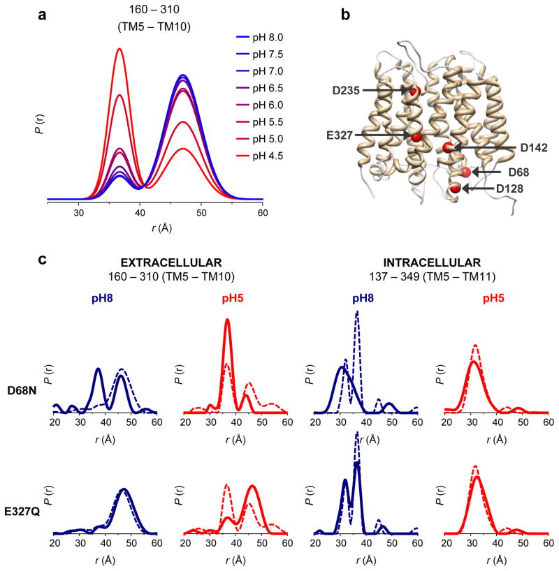
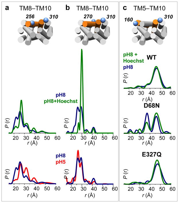
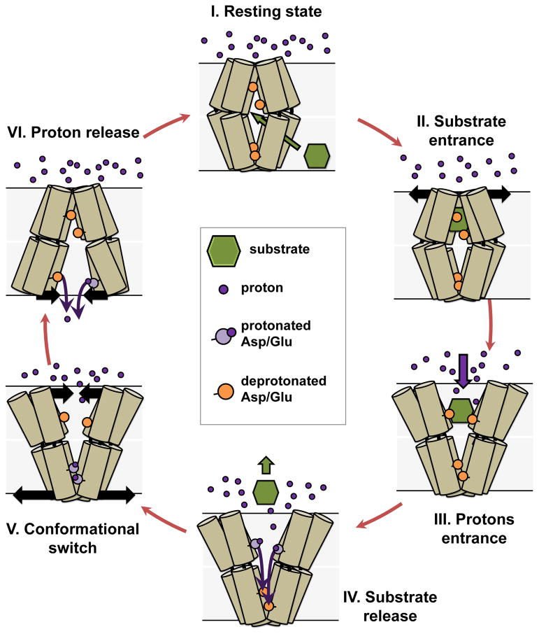
Multidrug antiporters of the major facilitator superfamily couple proton translocation to the extrusion of cytotoxic molecules. The conformational changes that underlie the transport cycle and the structural basis of coupling of these transporters have not been elucidated. Here we used extensive double electron-electron resonance measurements to uncover the conformational equilibrium of LmrP, a multidrug transporter from Lactococcus lactis, and to investigate how protons and ligands shift this equilibrium to enable transport. We find that the transporter switches between outward-open and outward-closed conformations, depending on the protonation states of specific acidic residues forming a transmembrane protonation relay. Our data can be framed in a model of transport wherein substrate binding initiates the transport cycle by opening the extracellular side. Subsequent protonation of membrane-embedded acidic residues induces substrate release to the extracellular side and triggers a cascade of conformational changes that concludes in proton release to the intracellular side.


Conflict of interest statement

Competing Financial Interests Statement

The authors declare no competing financial interests.

Figures

Figure 1
Figure 1. The extracellular side of LmrP closes at low pH
DEER distance distributions between labeled cysteine pairs located on the extracellular ends of TM helices. At pH 8 (blue curves) a single population (i.e. >70%) is observed for all distances. At pH 5 (red curves) a significant decrease in the distances is observed for pairs between the N-terminal (TMs 1–6) and C-terminal (TMs 7–12) bundles (panels a–h), while no effect is observed for distances within the same bundle (panels i–l). Distributions were normalized: r indicates interspin distance, P(r) indicates the distance probability. Residue numbers and positions of cysteine pairs are depicted on an LmrP model based on the crystal structure of the E. coli homolog EmrD (see Methods) viewed from the extracellular side. Targeted helices are highlighted in orange with TM numbers indicated atop. Asterisks denote peaks resulting from partial aggregation observed in some samples upon concentration.
Figure 2
Figure 2. Acidic pH opens the intracellular side of LmrP
DEER distance measurements on the intracellular side of LmrP, performed at pH 8 (blue curves) and pH 5 (red curves). Distances were measured either between the N-terminal and C-terminal bundles (panels a–f) or within the same bundle (panels g,h). Residue numbers and positions are depicted on an LmrP model based on the crystal structure of the E. coli homolog EmrD viewed from the intracellular side. Asterisks denote aggregation peaks.
Figure 3
Figure 3. The conformational equilibrium of LmrP is coupled to the protonation of acidic residues
(a) Distance distributions of the extracellular reporter pair L160C–I310C were determined at eight different pH values, ranging from 4.5 to 8.0 (color gradient from red to blue). To quantify the variation in population ratios as a function of pH, fits were carried out assuming a two-component Gaussian distance distribution in a home design software implemented in MATLAB (see Supplementary Fig. 7 and METHODS). At low pH, the short component (centered at 36 Å) dominates the bimodal distance distributions; at high pH the long component (centered at 47 Å) is dominant. (b) Key acidic residues depicted on an EmrD-based model of LmrP: Asp68 (bottom of TM2), Asp128 (bottom of TM4), Asp142 (center of TM5), Asp235 (top of TM7), Glu327 (center of TM10). (c) Protonation mimetic of key acidic residues can block the conformational switch. Single mutations D68N and E327Q were combined with double cysteine mutants L160C–I310C and V137C–S349C, which served as extracellular and intracellular reporters. Distance measurements at pH 5 (red curves) and pH 8 (blue curves) in the absence (dashed line) and presence (full line) of each mutation reveal the structural consequence of permanent protonation of these essential acidic residues.
Figure 4
Figure 4. Hoechst 33342 binding restricts TM8 conformational flexibility and stabilizes the outward-open conformation
(a–b) Distances distributions measured from the center (Cys270) and the extracellular end (I256C) of TM8 to the extracellular end of TM10 (I310C) at pH 8 (blue curves), pH 8 in the presence of 1 mM Hoechst 33342 (green curves) and pH 5 (red curves). The presence of substrate restricts the distributions to a single population, while TM8 adopts multiple conformations in apo LmrP, both at pH 5 and pH 8. (c) Effect of substrate binding (1 mM Hoechst 33342) on the extracellular conformation at pH 8 in the absence of a functional mutation; in combination with D68N; in combination with E327Q. The effect of the D68N mutation is partly reversed by the presence of the substrate.
Figure 5
Figure 5. Proposed LmrP transport cycle based on DEER distance measurements
For clarity, only six TMs of LmrP are depicted, with the extracellular side on top. The bilayer of the membrane is represented by shaded grey boxes and the transmembrane proton gradient illustrated by the large number of extracellular protons (purple spheres). (I) In the resting state, LmrP is occluded from the extracellular environment. The ligand (in green) enters from the membrane bilayer. (II) Substrate binding stabilizes the outward-open conformation. (III) The outward-open conformation exposes the binding site to extracellular protons (purple dots). (IV) Protons and/or water molecules enter the binding pocket and protonate carboxylic residues (orange circles, colored in light purple once protonated) that coordinate the substrate, thereby releasing it. (V) Protons access the acidic residues, eventually leading to protonation of D68, which induces closing of LmrP on the extracellular side and concomitant opening on the intracellular side. (VI) This opening will lead to (partial) exposure to the neutral intracellular milieu, allowing for deprotonation of Asp68 at which point the transporter resets to the resting state.

References

    1. Li XZ, Nikaido H. Efflux-mediated drug resistance in bacteria: an update. Drugs. 2009;69:1555–623. - PMC - PubMed
    1. Putman M, van Veen HW, Degener JE, Konings WN. The lactococcal secondary multidrug transporter LmrP confers resistance to lincosamides, macrolides, streptogramins and tetracyclines. Microbiology. 2001;147:2873–80. - PubMed
    1. Jardetzky O. Simple allosteric model for membrane pumps. Nature. 1966;211:969–70. - PubMed
    1. Tanford C. Mechanism of free energy coupling in active transport. Annu Rev Biochem. 1983;52:379–409. - PubMed
    1. Forrest LR, Krämer R, Ziegler C. The structural basis of secondary active transport mechanisms. Biochim Biophys Acta. 2011;1807:167–88. - PubMed

Publication types

LinkOut - more resources