. 2014 Jan;35(1):136-47.
doi: 10.1097/MAO.0000000000000003.

Prosthetic implantation of the human vestibular system

Affiliations

Prosthetic implantation of the human vestibular system

Justin S Golub et al. Otol Neurotol. 2014 Jan.

Abstract

Hypothesis: A functional vestibular prosthesis can be implanted in human such that electrical stimulation of each semicircular canal produces canal-specific eye movements while preserving vestibular and auditory function.

Background: A number of vestibular disorders could be treated with prosthetic stimulation of the vestibular end organs. We have previously demonstrated in rhesus monkeys that a vestibular neurostimulator, based on the Nucleus Freedom cochlear implant, can produce canal-specific electrically evoked eye movements while preserving auditory and vestibular function. An investigational device exemption has been obtained from the FDA to study the feasibility of treating uncontrolled Ménière's disease with the device.

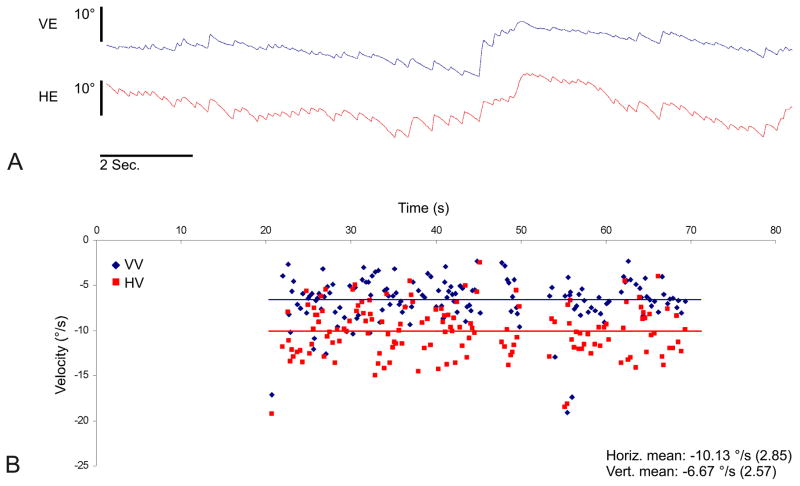
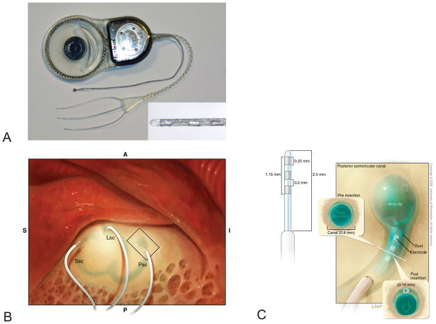
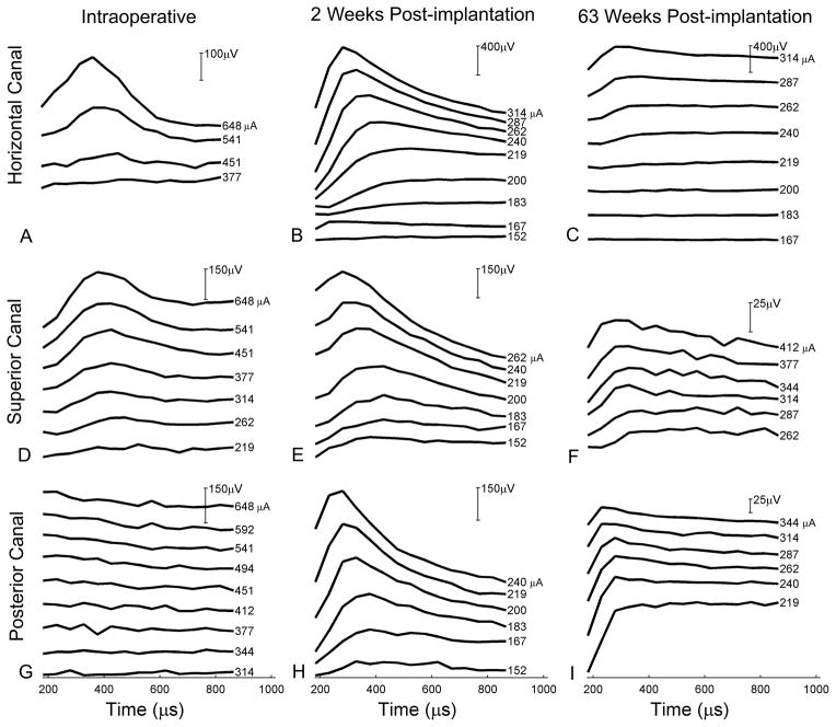
Methods: The UW/Nucleus vestibular implant was implanted in the perilymphatic space adjacent to the three semicircular canal ampullae of a human subject with uncontrolled Ménière's disease. Preoperative and postoperative vestibular and auditory function was assessed. Electrically evoked eye movements were measured at 2 time points postoperatively.

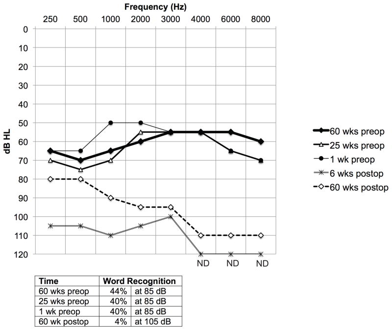
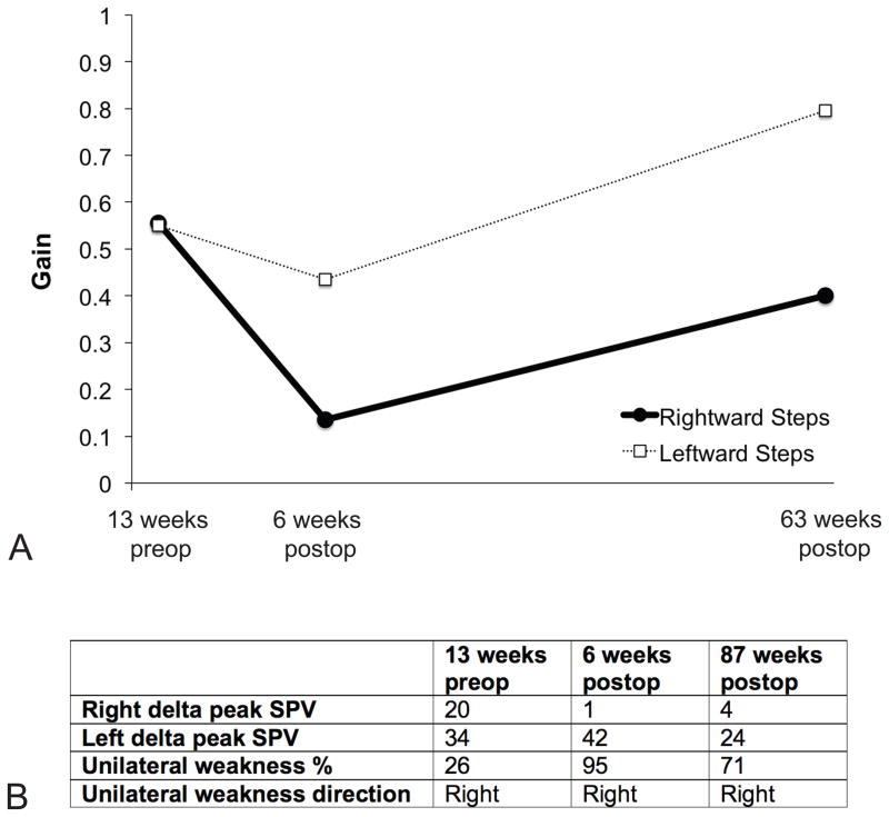
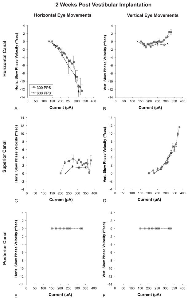
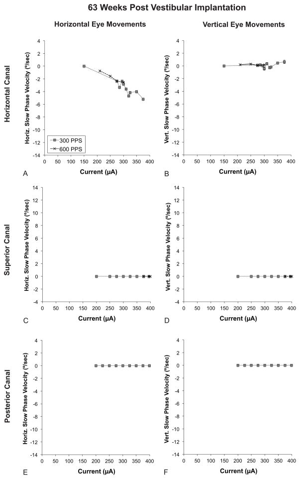
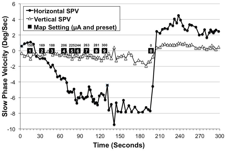
Results: Implantation of all semicircular canals was technically feasible. Horizontal canal and auditory function were largely, but not totally, lost. Electrode stimulation in 2 of 3 canals resulted in canal-appropriate eye movements. Over time, stimulation thresholds increased.

Conclusion: Prosthetic implantation of the semicircular canals in humans is technically feasible. Electrical stimulation resulted in canal-specific eye movements, although thresholds increased over time. Preservation of native auditory and vestibular function, previously observed in animals, was not demonstrated in a single subject with advanced Ménière's disease.


Figures

FIG. 1
FIG. 1
Videonystagmography of a patient with left-sided Ménière’s disease during an acute attack illustrating hypofunction of the ipsilateral vestibular system. A. Vertical (blue) and horizontal (red) eye position traces. B. Vertical (blue) and horizontal (red) slow-phase velocity (SPV) over time. Each point denotes the velocity of a single slow phase, while the lines represent the average slow-phase velocity over the course of the recording.
FIG. 2
FIG. 2
Design of the UW/Nucleus vestibular implant. A. Photograph displaying trifurcating electrode array with 3 electrodes per array (inset) as well as a ball ground electrode. The case serves as an additional ground electrode. B. Electrodes are implanted into the semicircular canal perilymphatic space adjacent to the ampulla. C. Enlargement of the box in B illustrating the theorized minimal distortion of the membranous labyrinth by the implanted electrode.
FIG. 3
FIG. 3
Audiometric data. Composite audiogram, including pure tone air conduction thresholds (above) and word recognition scores (below) for the implanted ear. Pre and postop refer to placement of the UW/Nucleus vestibular implant. ND = not detected.
FIG. 4
FIG. 4
Vestibular function data. A. Rotational chair velocity step testing. B. Caloric data. Delta peak SPV indicates the difference between the peak warm slow phase velocity and the peak cold slow phase velocity values. Note that the right ear was implanted. SPV = slow phase velocity.
FIG. 5
FIG. 5
Electrically evoked eye movements at an early time point (2 weeks post-implantation). All graphs depict slow phase velocity (SPV) versus current. Positive values denote either rightward or upward slow phase velocity. Refer to the inset in A for trace labels. A. Horizontal SPV during stimulation of the horizontal canal electrode. B. Vertical SPV during stimulation of the horizontal canal electrode. C. Horizontal SPV during stimulation of the superior canal electrode. D. Vertical SPV during stimulation of the superior canal electrode. E. Horizontal SPV during stimulation of the posterior canal electrode. F. Vertical SPV during stimulation of the posterior canal electrode. Note that the ranges of the X and Y-axes are the same for all graphs. Error bars represent SEM. PPS = pulses per second.
FIG. 6
FIG. 6
Electrically evoked eye movements at a late time point (63 weeks post-implantation). All graphs depict slow phase velocity (SPV) versus current. Positive values denote either rightward or upward slow phase velocity. Refer to the legend in part A for tracing labels. A. Horizontal SPV during stimulation of the horizontal canal electrode. B. Vertical SPV during stimulation of the horizontal canal electrode. C. Horizontal SPV during stimulation of the superior canal electrode. D. Vertical SPV during stimulation of the superior canal electrode. E. Horizontal SPV during stimulation of the posterior canal electrode. F. Vertical SPV during stimulation of the posterior canal electrode. The scale of the X and Y-axes is the same as in Fig. 5. Error bars represent SEM. PPS = pulses per second.
FIG. 7
FIG. 7
Electrically evoked compound action potentials (ECAPs). A. Horizontal canal intraop. B. Horizontal canal at 2 weeks postop. C. Horizontal canal at 63 weeks postop. D. Superior canal intraop. E. Superior canal at 2 weeks postop. F. Superior canal at 63 weeks postop. G. Posterior canal intraop. H. Posterior canal at 2 weeks postop. I. Posterior canal at 63 weeks postop. Note that the Y-axis scale varies (indicated by vertical bar at upper-right of graphs).
FIG. 8
FIG. 8
Slow phase velocity over time as “take-home” map settings are increased from “volume 1” (programmed to 150 μA) to “volume 9” (programmed to 300 μA). This simulates the sequence of stimuli the device would produce if the user were to titrate the prosthetic stimulation current to the intensity of a Ménière’s attack. The depicted simulation was performed in the laboratory in the absence of a Ménière’s attack at 6 weeks postoperatively. Positive values denote either rightward or upward slow phase velocity. Note that the subject’s actual take home map ranged from 150 μA to 350 μA.

References

    1. Gong W, Merfeld DM. Prototype neural semicircular canal prosthesis using patterned electrical stimulation. Ann Biomed Eng. 2000;28:572–81. - PubMed
    1. Della Santina C, Migliaccio A, Patel A. Electrical stimulation to restore vestibular function development of a 3-d vestibular prosthesis. Conf Proc IEEE Eng Med Biol Soc. 2005;7:7380–5. - PMC - PubMed
    1. Merfeld DM, Haburcakova C, Gong W, et al. Chronic vestibulo-ocular reflexes evoked by a vestibular prosthesis. IEEE Trans Biomed Eng. 2007;54:1005–15. - PubMed
    1. Phillips JO, Bierer SM, Ling L, et al. Real-time communication of head velocity and acceleration for an externally mounted vestibular prosthesis. Proceedings of the IEEE Engineering in Medicine and Biology Society. 2011;2011:3537–41. - PubMed
    1. Dai C, Fridman GY, Davidovics NS, et al. Restoration of 3D vestibular sensation in rhesus monkeys using a multichannel vestibular prosthesis. Hear Res. 2011;281:74–83. - PMC - PubMed

Publication types