

Okadaic acid toxin at sublethal dose produced cell proliferation in gastric and colon epithelial cell lines
- PMID: 24317467
- PMCID: PMC3877884
- DOI: 10.3390/md11124751
Okadaic acid toxin at sublethal dose produced cell proliferation in gastric and colon epithelial cell lines
Abstract
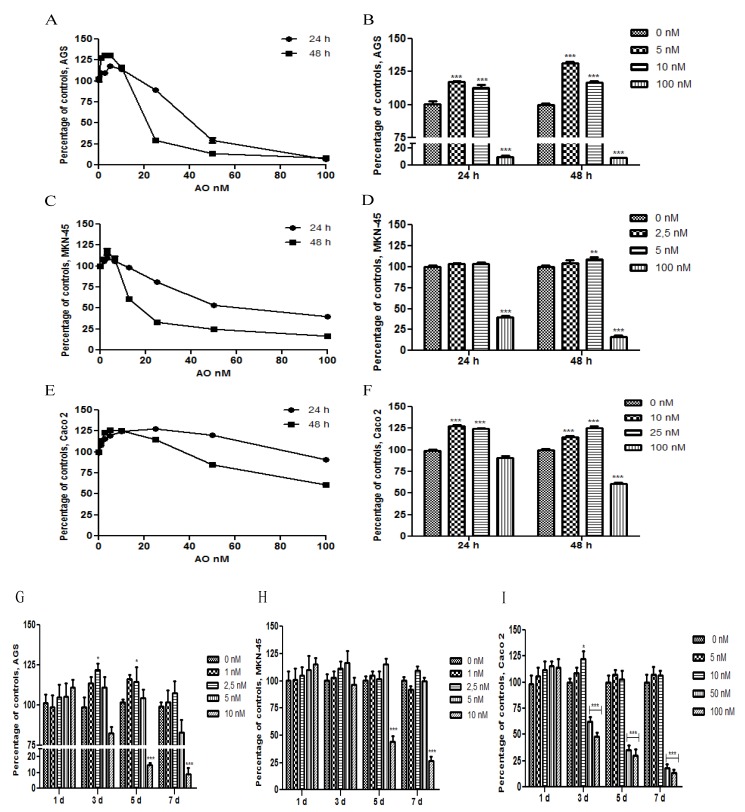
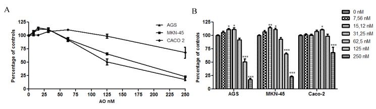
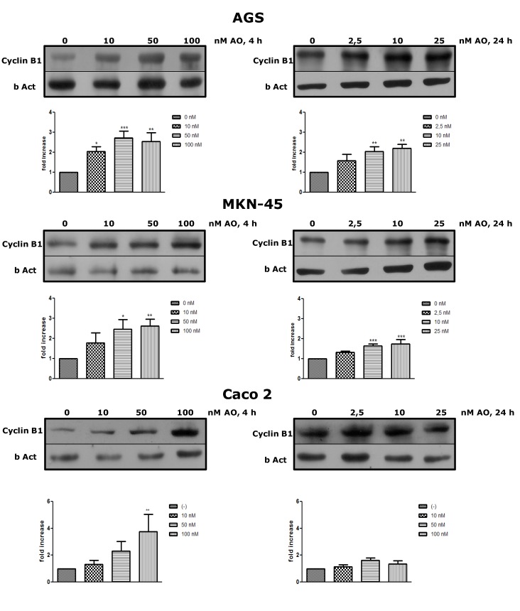
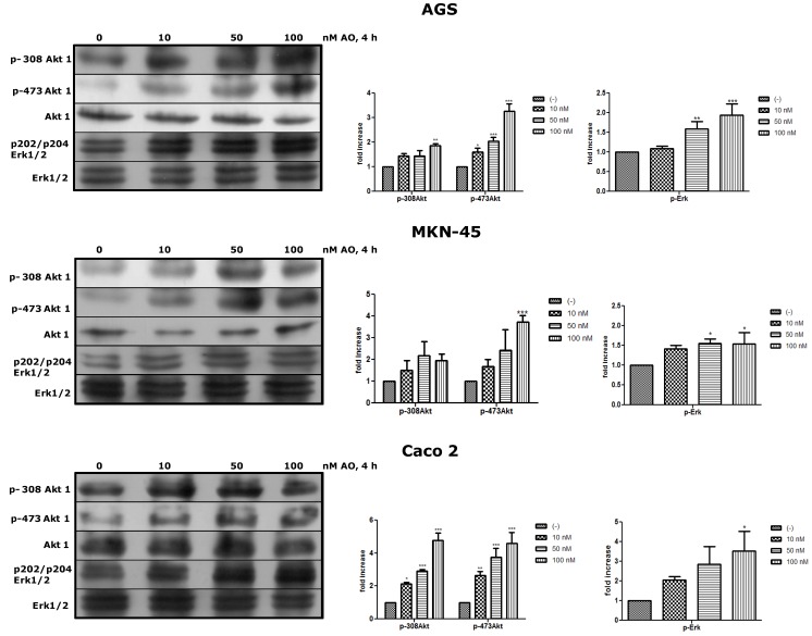
The aim of this study was to analyze the effect of Okadaic Acid (OA) on the proliferation of gastric and colon epithelial cells, the main target tissues of the toxin. We hypothesized that OA, at sublethal doses, activates multiple signaling pathways, such as Erk and Akt, through the inhibition of PP2A. To demonstrate this, we carried out curves of doses and time response against OA in AGS, MKN-45 and Caco 2 cell lines, and found an increase in the cell proliferation at sublethal doses, at 24 h or 48 h exposure. Indeed, cells can withstand high concentrations of the toxin at 4 h exposure, the time chosen considering the maximum time before total gastric emptying. We have proved that this increased proliferation is due to an overexpression of Cyclin B, a cyclin that promotes the passage from G2 to mitosis. In addition, we have demonstrated that OA induces activation of Akt and Erk in the three cells lines, showing that OA can activate pathways involved in oncogenesis. In conclusion, this study contributes to the knowledge about the possible effects of chronic OA consumption.
Figures
References
-
- Alexander J., Audunsson G.A., Benford D., Cockburn A., Cradevi J.P., Dogliotti E., Domenico A.D., Fernandez-Cruz M.L., Fink-Gremmels J., Furst P., et al. Marine biotoxins in shellfish—okadaic acid and analogues—Scientific Opinion of the Panel on Contaminants in the Food chain. EFSA J. 2008;589:1–62.
-
- Aune T., Stabell O.B., Nordstoga K., Tjotta K. Oral toxicity in mice of algal toxins from the diarrheic shellfish toxin (DST) complex and associated toxins. J. Nat. Toxins. 1998;7:141–158. - PubMed
Publication types
MeSH terms
Substances
LinkOut - more resources
Full Text Sources
Other Literature Sources
Molecular Biology Databases
Miscellaneous
