. 2014 Jan;21(1):43-8.
doi: 10.1038/nsmb.2721. Epub 2013 Dec 8.

Copper-transporting P-type ATPases use a unique ion-release pathway

Affiliations

Copper-transporting P-type ATPases use a unique ion-release pathway

Magnus Andersson et al. Nat Struct Mol Biol. 2014 Jan.

Abstract

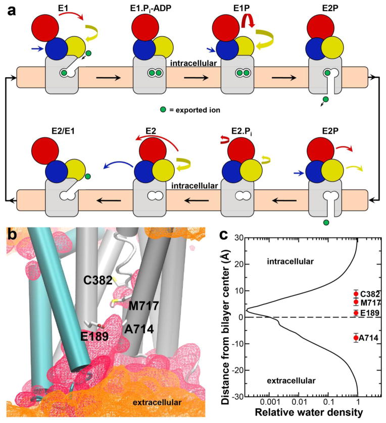
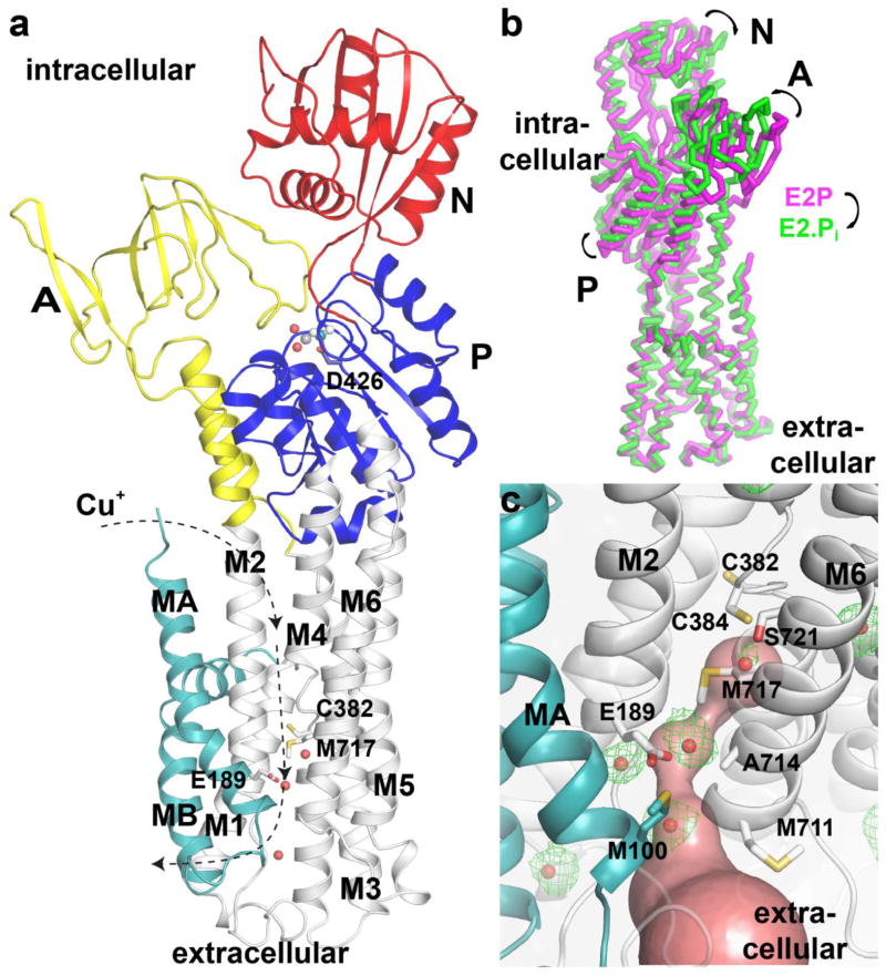
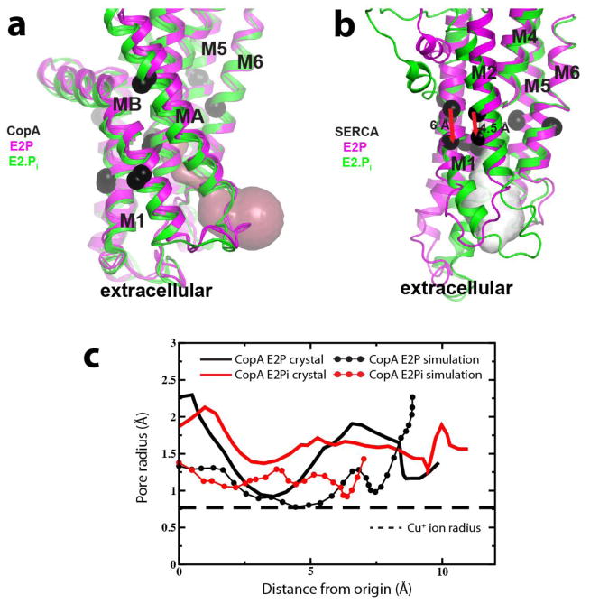
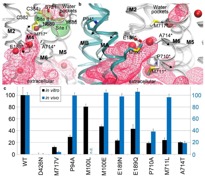
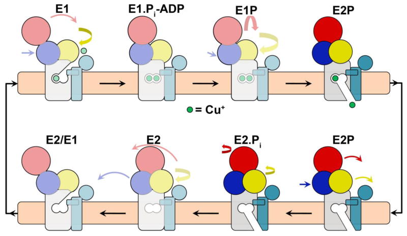
Heavy metals in cells are typically regulated by PIB-type ATPases. The first structure of the class, a Cu(+)-ATPase from Legionella pneumophila (LpCopA), outlined a copper transport pathway across the membrane, which was inferred to be occluded. Here we show by molecular dynamics simulations that extracellular water solvated the transmembrane (TM) domain, results indicative of a Cu(+)-release pathway. Furthermore, a new LpCopA crystal structure determined at 2.8-Å resolution, trapped in the preceding E2P state, delineated the same passage, and site-directed-mutagenesis activity assays support a functional role for the conduit. The structural similarities between the TM domains of the two conformations suggest that Cu(+)-ATPases couple dephosphorylation and ion extrusion differently than do the well-characterized PII-type ATPases. The ion pathway explains why certain Menkes' and Wilson's disease mutations impair protein function and points to a site for inhibitors targeting pathogens.


Conflict of interest statement

COMPETING FINANCIAL INTERESTS

The authors declare no competing financial interests.

Figures

Figure 1
Figure 1. MD simulations suggest the E2.Pi state to be open in CopA
a, Schematics of the classical P-type ATPase reaction cycle, known e.g. for Ca2+-transporting SERCA. The intracellular A-, P- and N-domains are colored yellow, blue and red, respectively, while the M-domain is gray. Ions (two Ca2+ for SERCA, shown in green) are transported accompanied by phosphate hydrolysis and structural rearrangements (marked by arrows). Note that the transmembrane domain occludes upon initiation of dephosphorylation (E2.Pi). b, Average representation from the MD simulation of the CopA E2.Pi state (pdb-id: 3RFU). The transmembrane domain is shown with helices MA-MB (Class-IB specific) and M1-M6 depicted in cyan and grey, respectively. The Cu+-binding residues Cys382 and Met717 as well as Glu189 and Ala714Thr at the exit pathway are shown as sticks. Lipid phosphates and water are shown as orange and red density surfaces at 5 % and 20 % occupancies, respectively (the fraction of presence in simulation frames). Water solvation reaches the ion binding residues. c, Density plot for the water distribution of the E2.Pi MD simulation showing the number of water molecules relative to bulk solution along the membrane normal within 7 Å from the protein (intracellular side positive). The centers-of-mass with corresponding error bars are depicted for Cys382, Met717, Glu189 and Ala714. Cu+ must pass more than half of the membrane from the intramembanous ion-binding residues Cys382 and Met717 to be released to the extracellular side.
Figure 2
Figure 2. Crystal waters of the E2-BeF3 structure support the copper release pathway
The domains are colored as in Fig. 1a. a, The E2-BeF3 structure determined in this work. Black arrows mark the Cu+ transport direction. Crystal waters located in close vicinity to Cys382, Met717, Glu189 and Ala714 are shown as red spheres. b, Superimpositions of the TM domains of the E2P (purple) and E2.Pi (green) states of LpCopA. Intracellular domains change their configuration from the E2P to the E2.Pi state (black arrows) while the TM domains remain rigid. c, Close-up of the extrusion pathway with residual Fobs-Fcalc electron density (green mesh at 2.5σ, prior to modeling) of BeF3-bound LpCopA with key residues labeled. The opening from the Cu+ high-affinity coordinating residues Cys382, Cys384 and Met717 found in the E2P conformation found using the software CAVER is shown as a red surface and overlays with crystallographic water molecules shown as red spheres.
Figure 3
Figure 3. The CopA release pathway is unique and able to accommodate Cu+ ions
a, View of the transmembrane domains of the E2P (purple) and E2.Pi (green) structures of SERCA and its release pathway (white). Black spheres indicate the movements of one Cα atom in each helix and transmembrane helices M7-M10 have been removed for clarity. Significant shifts of helices M1-M2 and M3-M4 occlude SERCA in the E2P to E2.Pi. See also Supplementary Fig. 4a. b, Equivalent view as in panel d for CopA with its release pathway (purple) showing that CopA remains rigid in the E2P to E2.Pi transition. See also Supplementary Fig. 4b. c, Pore radii of the E2P and E2.Pi crystal structures and average structures from last 10 ns of the corresponding simulations compared to the size of Cu+. The origin was set close to the centers-of-mass of the internal Cu+ coordinators Cys382, Cys384 and Met717. See Supplementary Fig. 10 for methods specification and corresponding SERCA analyses.
Figure 4
Figure 4. Copper transfer from the high-affinity binding sites and the extracellular release pathway
Hydration and sidechain dynamics of the LpCopA crystal structures colored as in Fig. 1a and with E2P crystal waters shown as solid, red spheres. Residues included in the functional analysis summarized in Fig. 4c are marked by asterisks. a, The E2P state simulation. Crystallographic and simulated (averaged over the last 20 ns) sidechain configurations are shown for Met717 and Glu189, while the E2:BeF3 crystal structure represents Cys382, Cys384, Tyr688, Asn689, Ala714 and Ser721 as they do not rearrange significantly during simulations. Green transparent spheres represent the approximate locations of calcium sites of SERCA (pdb-id: 3BA6), with Site I encircled in green, compared to the equivalent water pockets (shown as in Fig. 1a and encircled in red). Note that a bulk water pathway associates Glu189 with the extracellular environment. The intrinsic flexibility of Met717 and Glu189 suggests a shuttling role in Cu+-transfer. See also Supplementary Fig. 8. b, An average from the last 20 ns of the E2.Pi simulation (transparent) superimposed on the E2. AlF4 (pdb-id: 3RFU) crystal structure (solid), highlighting the opening in CopA, which involves the class-specific helix MA and common P-type ATPase core helices M2 and M6 and emerges from Pro94 and Pro710. Arrows mark movements of Cα atoms in MA and M6 (shown as cyan and grey spheres, analysis described in Supplementary Fig. 16). The red circle is equivalent to the one in panel a. c, Relative in vitro activity of LpCopA copper release pathway mutants as compared to wild-type (WT) protein and the inactive Asp426Asn mutant (black). Relative in vivo susceptibility for copper of the same mutations (blue, see also Supplementary Fig. 19), except for M100L, which was not cloned for the in vivo assay.
Figure 5
Figure 5. Mechanistic implications of the open E2.Pi state
a) The CopA domains are colored as in Fig. 1a and colored arrows indicate the coming movements of the corresponding domain to the following conformation. In contrast to the well-characterized Ca2+-P-type ATPase SERCA, no major movements are identified between the transmembrane domains of the E2P and E2.Pi LpCopA states, indicating a class-specific transport mechanism with an open release pathway associated with dephosphorylation.

References

    1. Lutsenko S, Kaplan JH. Organization of P-type ATPases: significance of structural diversity. Biochemistry. 1995;34:15607–13. - PubMed
    1. Kuhlbrandt W. Biology, structure and mechanism of P-type ATPases. Nat Rev Mol Cell Biol. 2004;5:282–95. - PubMed
    1. Arguello JM, Eren E, Gonzalez-Guerrero M. The structure and function of heavy metal transport P1B-ATPases. Biometals. 2007;20:233–48. - PubMed
    1. Vulpe C, Levinson B, Whitney S, Packman S, Gitschier J. Isolation of a candidate gene for Menkes disease and evidence that it encodes a copper-transporting ATPase. Nat Genet. 1993;3:7–13. - PubMed
    1. Bull PC, Thomas GR, Rommens JM, Forbes JR, Cox DW. The Wilson disease gene is a putative copper transporting P-type ATPase similar to the Menkes gene. Nat Genet. 1993;5:327–37. - PubMed

Publication types

MeSH terms

Associated data