

Proposal for a new mediastinal compartment classification of transverse plane images according to the Japanese Association for Research on the Thymus (JART) General Rules for the Study of Mediastinal Tumors
- PMID: 24317723
- PMCID: PMC3896522
- DOI: 10.3892/or.2013.2904
Proposal for a new mediastinal compartment classification of transverse plane images according to the Japanese Association for Research on the Thymus (JART) General Rules for the Study of Mediastinal Tumors
Abstract
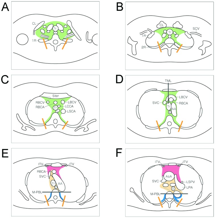
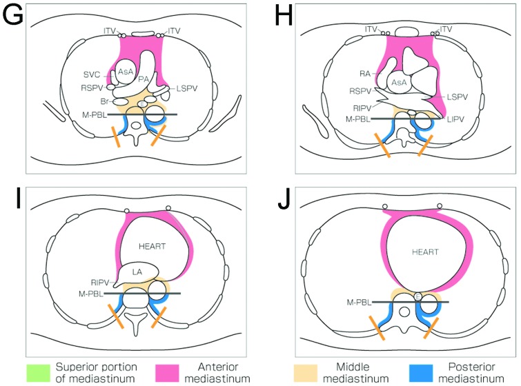
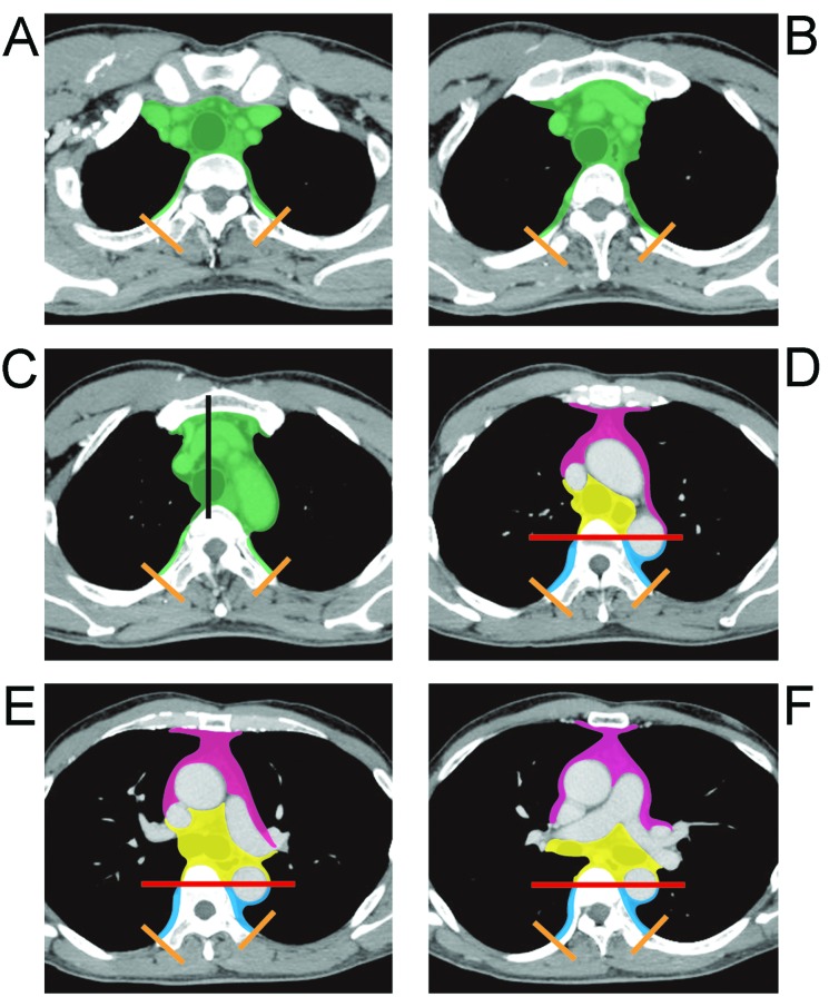
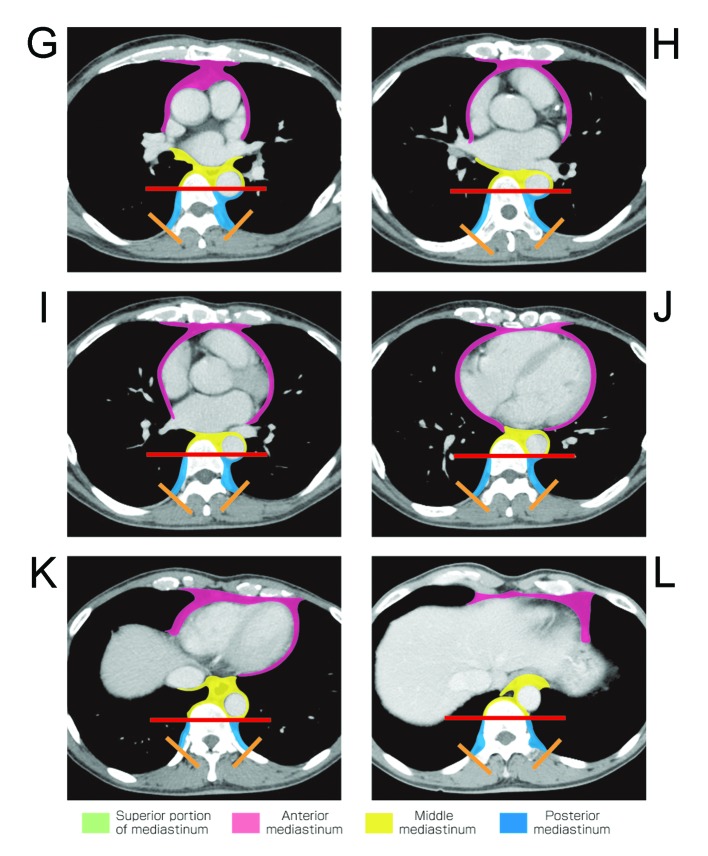
There is no existing worldwide published method for mediastinum compartment classification based on transverse section images for the differential diagnosis of mediastinal tumors. Herein, we describe a new method for anatomic mediastinal compartment classification using transverse section computed tomography (CT) images and the use of this method to classify mediastinal lesions, and thus evaluate whether the method is sufficiently user-friendly and useful. In a publication of the Japanese Association for Research on the Thymus (JART), we proposed the following four mediastinal compartments based on transverse CT images: superior portion of mediastinum, anterior mediastinum (prevascular zone), middle mediastinum (peri-tracheoesophageal zone), and posterior mediastinum (paravertebral zone). In the present study, we retrospectively analyzed 445 pathologically proven mediastinal mass lesions, and categorized them into the proposed four compartments by consensus reading. Mass lesions were classified into compartments based on the location of the lesion centroid, and each lesion was satisfactorily categorized into a compartment. Almost all thymic epithelial tumors (99%, 244/246), all 24 thymic malignant lymphomas and a majority of germ cell neoplasms (93%, 54/58) were classified as being in the anterior mediastinum compartment. The majority of intrathoracic goiters (82%, 14/17) were categorized as being in the superior portion of the mediastinum compartment. Approximately two-thirds of mass lesions in the middle mediastinum were cysts, including foregut and pericardial cysts. Approximately 80% of 37 mass lesions in the posterior mediastinum were neurogenic tumors. Correspondingly, 29 of the 49 neurogenic tumors (60%) were categorized as being in the posterior mediastinum, while 10 (20%) were in the superior portion of the mediastinum, 4 (8%) in the anterior mediastinum, and 6 (12%) in the middle mediastinum. Our findings showed that the newly proposed mediastinal compartment classification using transverse images appears to be user-friendly enough for practical clinical application and may be helpful in differential diagnoses.
Figures
References
-
- Gatzoulis MA. Mediastinum. In: Standring S, editor. Gray’s Anatomy. The Anatomical Basis of Clinical Practice. 40th edition. Churchill Livingstone (Elsevier); Philadelphia, PA: 2008. pp. 939–957.
-
- Fraser RS, Müller NL, Colman N, Paré PD. Fraser and Paré’s Diagnosis of Diseases of the Chest. 4th edition. WB Saunders; Philadelphia, PA: 1999. The mediastinum; pp. 196–234.
-
- Fraser RG, Paré JA. Diagnosis of Diseases of the Chest. 2nd edition. WB Saunders; Philadelphia, PA: 1977. The normal chest; pp. 1–183.
-
- Felson B. Chest Roentgenology. WB Saunders; Philadelphia, PA: 1973.
-
- Heitzman ER. The Mediastinum. 2nd edition. Springer-Verlag; New York: 1988.
Publication types
MeSH terms
Supplementary concepts
LinkOut - more resources
Full Text Sources
Other Literature Sources
