
Hippocampal sclerosis after febrile status epilepticus: the FEBSTAT study

Darrell V Lewis et al. Ann Neurol. 2014 Feb.

Abstract

Objective: Whether febrile status epilepticus (FSE) produces hippocampal sclerosis (HS) and temporal lobe epilepsy (TLE) has long been debated. Our objective is to determine whether FSE produces acute hippocampal injury that evolves to HS.

Methods: FEBSTAT and 2 affiliated studies prospectively recruited 226 children aged 1 month to 6 years with FSE and controls with simple febrile seizures. All had acute magnetic resonance imaging (MRI), and follow-up MRI was obtained approximately 1 year later in the majority. Visual interpretation by 2 neuroradiologists informed only of subject age was augmented by hippocampal volumetrics, analysis of the intrahippocampal distribution of T2 signal, and apparent diffusion coefficients.

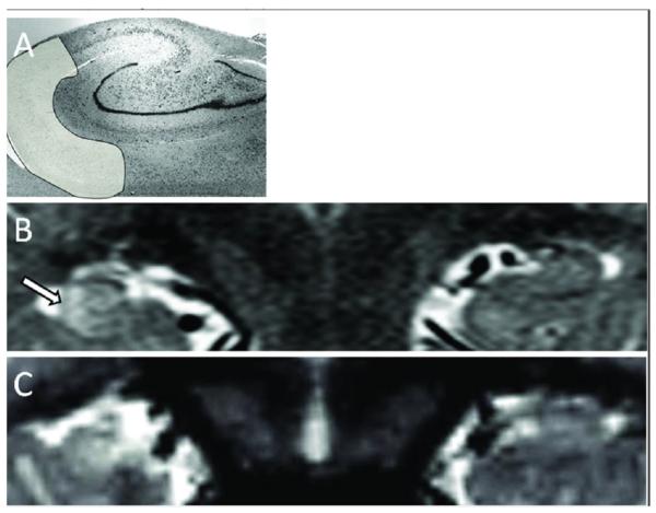
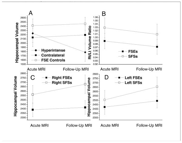
Results: Hippocampal T2 hyperintensity, maximum in Sommer's sector, occurred acutely after FSE in 22 of 226 children in association with increased volume. Follow-up MRI obtained on 14 of the 22 with acute T2 hyperintensity showed HS in 10 and reduced hippocampal volume in 12. In contrast, follow-up of 116 children without acute hyperintensity showed abnormal T2 signal in only 1 (following another episode of FSE). Furthermore, compared to controls with simple febrile seizures, FSE subjects with normal acute MRI had abnormally low right to left hippocampal volume ratios, smaller hippocampi initially, and reduced hippocampal growth.

Interpretation: Hippocampal T2 hyperintensity after FSE represents acute injury often evolving to a radiological appearance of HS after 1 year. Furthermore, impaired growth of normal-appearing hippocampi after FSE suggests subtle injury even in the absence of T2 hyperintensity. Longer follow-up is needed to determine the relationship of these findings to TLE.


Figures

Figure 1
Figure 1
Increased Hippocampal T2 Signal Following FSE. A) Nissl stain of hippocampal body outlining area of Sommer's sector (Courtesy G. Mathern). B) Acutely swollen and hyperintense right hippocampus of a 13 month old male 3 days after a 120 min long episode of FSE. Note the right hippocampus is larger and has increased T2 signal most prominent in Sommer's sector (Arrow). C) Follow up MRI 6 months later of same child showing the right hippocampus now smaller and the T2 signal increase is persistent but no longer maximum in Sommer's sector.
Figure 2
Figure 2
Increased T2 on Acute and Follow-Up MRIs. Inset - ROIs overlying hippocampal sectors, SS = Sommer's Sector, Sub =subiculum. Plot shows increase in T2 intensity by hippocampal sector in the acute (filled squares; N=21) and follow-up (open squares; N=13) MRIs. On acute scans, the mean (± St Dev) increase in T2 signal across all three Sommer's sector ROIs was 0.141 (± 0.136) vs. 0.057 (±0.099) for all other sectors (t-test; p<0.001). Solid and dashed lines = means over SS and Non-SS sectors at Acute and Follow-Up, respectively. Bars = 95% Confidence Intervals.
Figure 3
Figure 3
Hippocampal Volumes from Acute and Follow-up MRIs. A) Hyperintense hippocampi (Hyperintense; N=15) vs. hippocampi contralateral to them (Contralateral; N=13) and control hippocampi from FSE subjects with normal acute MRIs (FSE Controls; N=30). The number of hyperintense hippocampi is greater than the number of contralateral hippocampi because one subject had bilateral hyperintensity. Note that the hyperintense hippocampi lost approximately 20% of volume between the initial and follow up MRIs, whereas the contralateral and FSE control hippocampal volumes increased. The mixed linear model indicated that, at follow-up MRI. the mean volume of hyperintense hippocampi was less than the controls (p<0.0001) and the contralaterals (p=0.0001). At the baseline MRI, the mean volumes of the groups were not significantly different. B) Rt/Lt volume ratios of FSE subjects with normal acute MRIs were less than SFS subjects with normal acute MRIs (mean estimated Rt/Lt volume ratios of 0.98 vs. 1.03 respectively; F-test p=0.02). Rt/Lt ratios did not change significantly from baseline to follow-up MRIs (F test p=0.10). C) Right hippocampal volumes of FSE were less than SFS group by 171 and 262 mm3 at the acute and follow-up MRIs respectively (Bonferroni adjusted p=0.03 for acute and p <0.001 for follow-up). D) Left hippocampal volumes of FSE were less than SFS subjects by 82 and 162 mm3 at the acute and follow-up MRIs although these differences did not attain significance (Bonferroni adjusted p=0. 90 for acute and p= 0.07 for follow-up). From the acute to follow-up MRI, the volumes of both sides increased for SFS subjects (Bonferroni adjusted left side: p = 0.03; right side: p = 0.11). For FSE subjects, the slope of growth was flatter and volume change was not significant on either side (left side: p = 0.74; right side: p = 1.00). Bars are 95% confidence intervals. For B, C and D the groups were identical with FSEs (N=59) and SFS controls (N=38).

Comment in

  • Reply: To PMID 24318290.
    Lewis D, Hesdorffer D, Moshé SL, Shinnar S; FEBSTAT Study Team. Lewis D, et al. Ann Neurol. 2014 Aug;76(2):316-7. doi: 10.1002/ana.24206. Epub 2014 Jul 16. Ann Neurol. 2014. PMID: 24961408 No abstract available.
  • Hippocampal sclerosis and other forms of status epilepticus.
    Yoong M, Martinos MM, Pujar S, Scott RC, Chin RF. Yoong M, et al. Ann Neurol. 2014 Aug;76(2):316. doi: 10.1002/ana.24207. Epub 2014 Jul 9. Ann Neurol. 2014. PMID: 24961513 No abstract available.

References

    1. Febrile Seizures. National Institute of Health; Bethesda, MD: 1980. Febrile seizures: Consensus development conference summary.
    1. Shinnar S. Febrile Seizures and Mesial Temporal Sclerosis. Epilepsy Curr. 2003;3:115–8. - PMC - PubMed
    1. Hesdorffer DC, Benn EK, Bagiella E, et al. Distribution of febrile seizure duration and associations with development. AnnNeurol. 2011;70:93–100. - PMC - PubMed
    1. Shinnar S, Pellock JM, Moshe SL, et al. In whom does status epilepticus occur: age-related differences in children. Epilepsia. 1997;38:907–14. - PubMed
    1. Mathern GW, Adelson PD, Cahan LD, Leite JP. Hippocampal neuron damage in human epilepsy: Meyer's hypothesis revisited. ProgBrain Res. 2002;135:237–51. - PubMed

Publication types