. 2013 Dec;4(12):2462-75.
doi: 10.18632/oncotarget.1578.

High aldehyde dehydrogenase activity identifies cancer stem cells in human cervical cancer

Affiliations

High aldehyde dehydrogenase activity identifies cancer stem cells in human cervical cancer

Shu-Yan Liu et al. Oncotarget. 2013 Dec.

Abstract

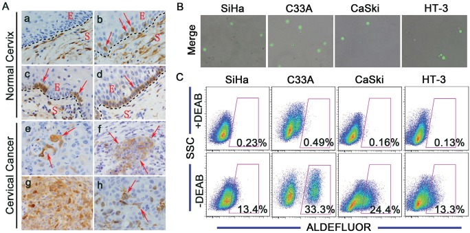
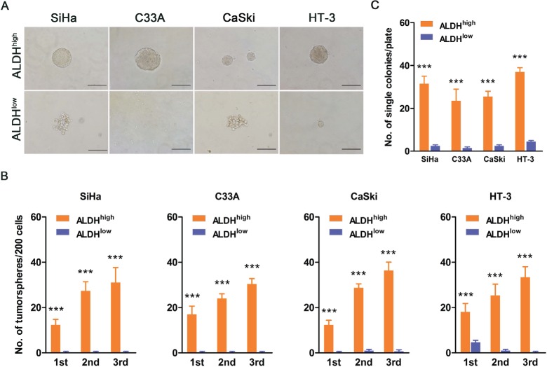
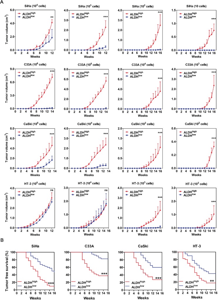
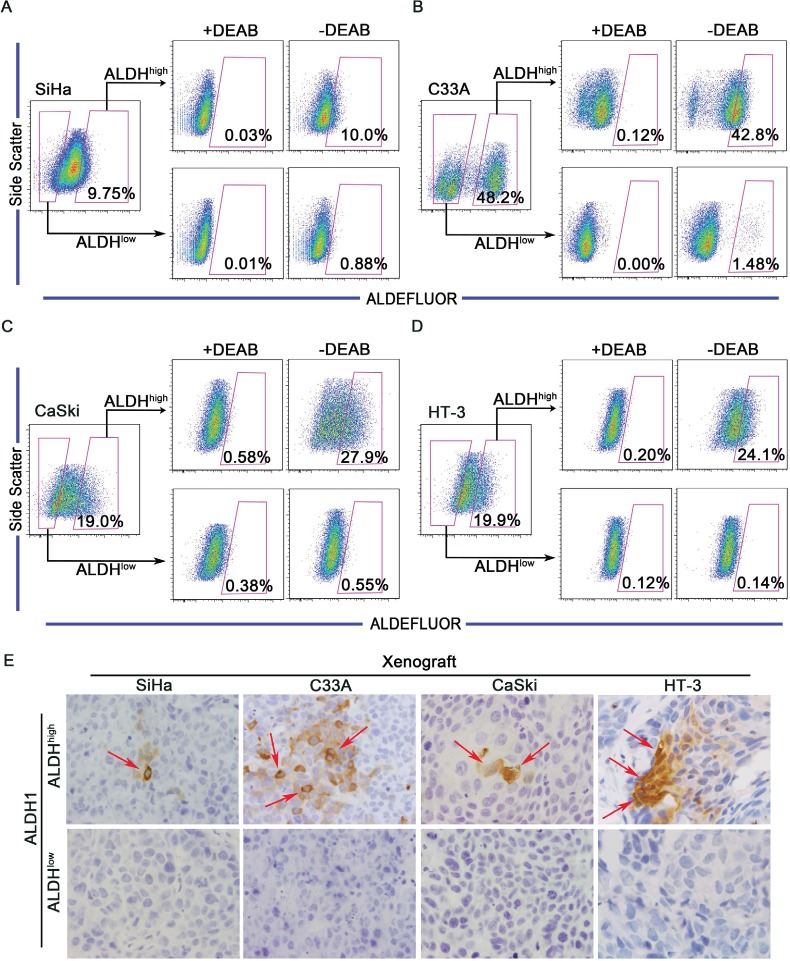
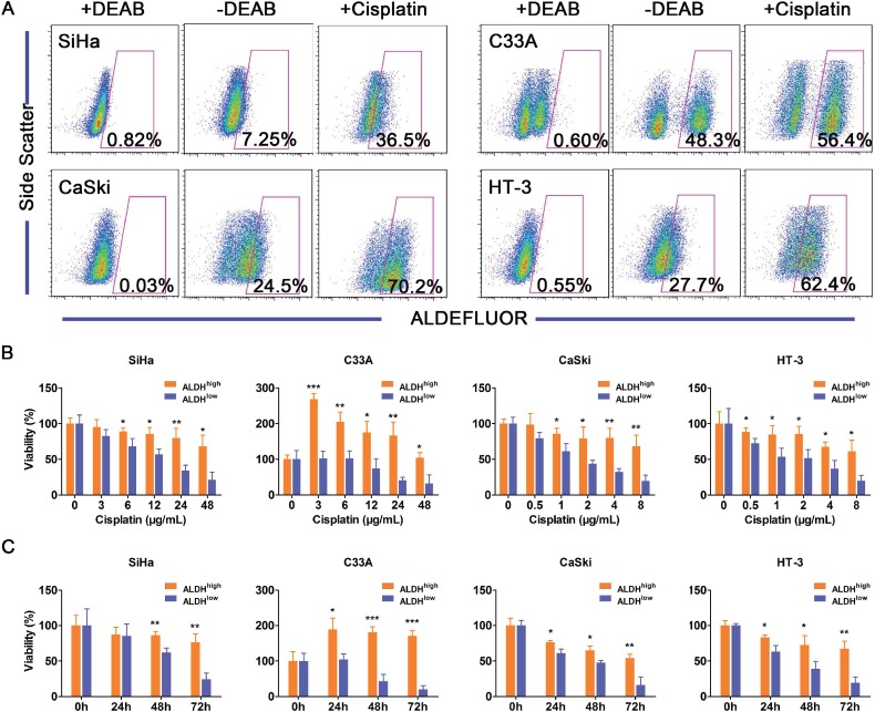
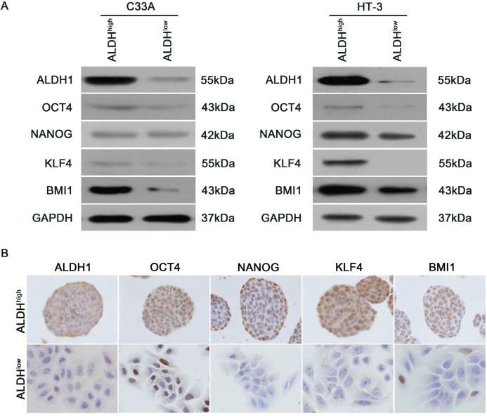
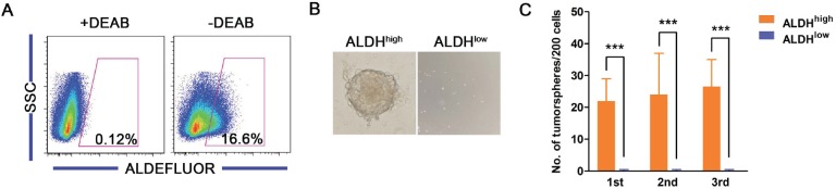
High aldehyde dehydrogenase (ALDH) activity characterizes a subpopulation of cells with cancer stem cell (CSC) properties in several malignancies. To clarify whether ALDH can be used as a marker of cervical cancer stem cells (CCSCs), ALDH high and ALDH low cells were sorted from 4 cervical cancer cell lines and 5 primary tumor xenografts and examined for CSC characteristics. Here, we demonstrate that cervical cancer cells with high ALDH activity fulfill the functional criteria for CSCs: (1) ALDH high cells, unlike ALDH low cells, are highly tumorigenic in vivo; (2) ALDH high cells can give rise to both ALDH high and ALDH low cells in vitro and in vivo, thereby establishing a cellular hierarchy; and (3) ALDH high cells have enhanced self-renewal and differentiation potentials. Additionally, ALDH high cervical cancer cells are more resistant to cisplatin treatment than ALDH low cells. Finally, expression of the stem cell self-renewal-associated transcription factors OCT4, NANOG, KLF4 and BMI1 is elevated in ALDH high cervical cancer cells. Taken together, our data indicated that high ALDH activity may represent both a functional marker for CCSCs and a target for novel cervical cancer therapies.


Figures

Figure 1
Figure 1. ALDH expression in human cervical tissue specimens and cervical cancer cell lines
A, Representative photos of immunostained specimens showing ALDH1 expression in normal cervical (a-d) and cervical cancer (e-h) tissues. a, no ALDH1-positive cells; b, dot-scattered ALDH1 expression; c, focal distribution of ALDH1-positive cells; d, all basal cells of the normal cervix are ALDH1-positive; e, dot-scattered ALDH1 expression; f, focal distribution of ALDH1-positive cells; g, diffuse ALDH1 expression; h, ALDH1-positive cells in xenograft tissue from mice. Red arrows indicate ALDH1-positive cells. S, Stroma; E, Epithelium; Magnifications, 1000×. B, Cells were labeled using the ALDEFLUOR kit, and ALDHhigh cells (bright green fluorescence) were detected by fluorescent microscopy. C, ALDH enzyme activity in 4 cervical cancer cell lines was analyzed by flow cytometry. As a negative control, cells were treated with the specific ALDH inhibitor DEAB. The gated cells represent the ALDHhigh cells.
Figure 2
Figure 2. ALDHhigh cervical cancer cells exhibit enhanced self-renewal capacity
A, Representative photos of tumorspheres formed by ALDHhigh and ALDHlow cells are shown. Bar, 200 µm. B, The number of tumorspheres/200 cells was counted from 3 consecutive passages. C, The number of wells containing tumorspheres was counted. ***, p<0.001. Data represent mean ± SD of triplicate experiments.
Figure 3
Figure 3. Tumorigencity of ALDHhigh and ALDHlow cells from 4 cervical cancer cell lines in NOD/SCID mice
A, The volume of xenograft tumors formed by different numbers of ALDHhigh and ALDHlow cervical cancer cells was monitored over time. B, Kaplan-Meier plots showing the tumor-free survival after injection. ns, not significant; *, p<0.05; **, p<0.01; ***, p<0.001. Data represent mean ± SD of tumor volumes at different time points of 10 mice in each group.
Figure 4
Figure 4. ALDHhigh cervical cancer cells are capable of differentiating in vitro and in vivo
A-D, ALDHhigh and ALDHlow cells were isolated from the SiHa (A), C33A (B), CaSki (C) or HT-3 (D) cell lines and cultured in DMEM medium supplemented with 10% FBS for 2 weeks. The ALDH enzyme activity was then analyzed by flow cytometry. Cells treated with DEAB served as a negative control. The gated cells represent the ALDHhigh cells. E, Expression of ALDH1 was detected by IHC in xenograft tumors from ALDHhigh and ALDHlow cells. Red arrows indicate ALDH1-positive cells
Figure 5
Figure 5. ALDHhigh cells are more resistant to cisplatin than ALDHlow cells
A, The ALDH activity of the cervical cancer cell lines was analyzed after exposure to cisplatin (3 µg/mL for SiHa and C33A cells or 1 µg/mL for CaSki and HT-3 cells) for 3 days. The percentage of ALDHhigh cells in the 4 cervical cancer cell lines was analyzed by flow cytometry following exposure to cisplatin for 3 days. B, Cell viability of the ALDHhigh and ALDHlow cervical cancer cells was measured using an MTT assay after treatment with different concentrations of cisplatin for 24 h. C, Cell viability of the ALDHhigh and the ALDHlow cervical cancer cells was measured using an MTT assay after treatment with a constant dose of cisplatin for 0, 24, 48 or 72 h. *, p<0.05; **, p<0.01; ***, p<0.001. Data represent mean ± SD of triplicate experiments.
Figure 6
Figure 6. Expression of stem cell-associated markers in ALDHhigh and ALDHlow cells
A, Western blot analysis of the protein levels of the stem cell-associated transcription factors OCT4, NANOG, KLF4 and BMI1 in the ALDHhigh and ALDHlow subpopulations from C33A and HT-3 cells. GAPDH was used as a loading control. B, The expression of stem cell-associated transcription factors in tumorspheres formed by ALDHhigh and ALDHlow SiHa cells was measured by IHC analysis.
Figure 7
Figure 7. Characteristics of ALDHhigh cells from primary cervical cancers
A, The ALDH enzyme activity of primary cervical cancer cells was analyzed by flow cytometry. B, Representative photos of tumorspheres formed by ALDHhigh and ALDHlow cells sorted from primary cervical cancer are shown. C, The number of tumorspheres formed by ALDHhigh and ALDHlow cells isolated from the primary cervical cancer was counted from 3 consecutive passages. ***, p<0.001. Data represent mean ± SD of triplicate experiments.

References

    1. Jemal A, Bray F, Center MM, Ferlay J, Ward E, Forman D. Global cancer statistics. CA Cancer J Clin. 2011;61(2):69–90. - PubMed
    1. Ferlay J, Shin HR, Bray F, Forman D, Mathers C, Parkin DM. Estimates of worldwide burden of cancer in 2008: GLOBOCAN 2008. Int J Cancer. 2010;127(12):2893–2917. - PubMed
    1. Walboomers JM, Jacobs MV, Manos MM, Bosch FX, Kummer JA, Shah KV, Snijders PJ, Peto J, Meijer CJ, Munoz N. Human papillomavirus is a necessary cause of invasive cervical cancer worldwide. J Pathol. 1999;189(1):12–19. - PubMed
    1. Bosch FX, Lorincz A, Munoz N, Meijer CJ, Shah KV. The causal relation between human papillomavirus and cervical cancer. J Clin Pathol. 2002;55(4):244–265. - PMC - PubMed
    1. Carter JR, Ding Z, Rose BR. HPV infection and cervical disease: a review. Aust N Z J Obstet Gynaecol. 2011;51(2):103–108. - PubMed

Publication types