

Atypical, abscessated nasopharyngeal polyp associated with expansion and lysis of the tympanic bulla
- PMID: 24319059
- PMCID: PMC11164163
- DOI: 10.1177/1098612X13514421
Atypical, abscessated nasopharyngeal polyp associated with expansion and lysis of the tympanic bulla
Abstract
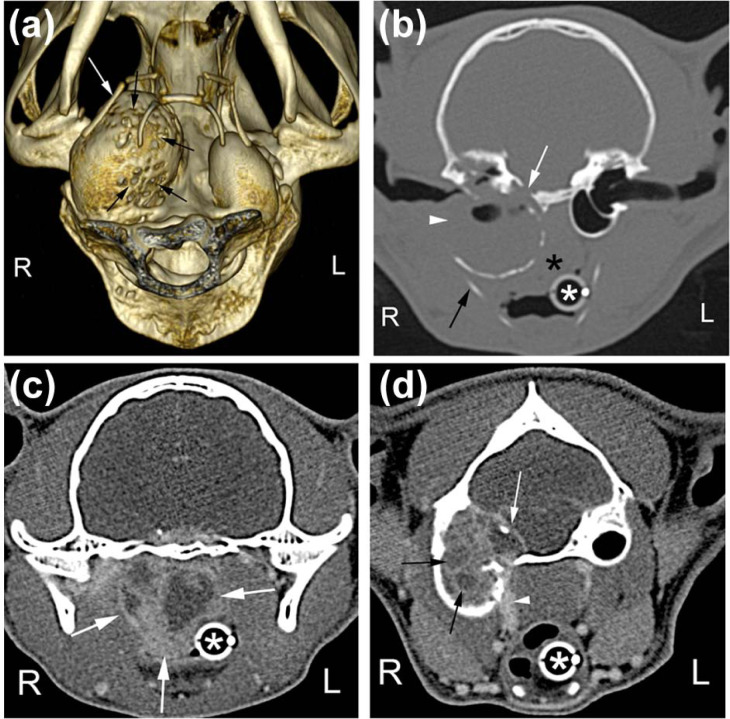
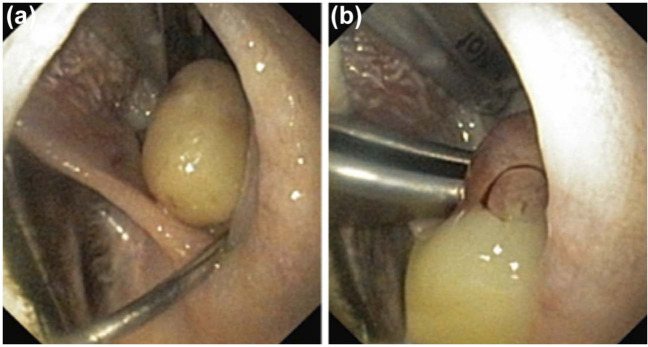
A 5-year-old, male neutered domestic shorthair cat was referred for investigation of lethargy, weight loss, pyrexia and upper respiratory tract signs. On computed tomography, an expansile, osteodestructive lesion in the right tympanic bulla was identified. A soft tissue mass extended from the bulla into the nasopharynx, cranium and subcutaneous tissues. The nasopharyngeal mass ruptured during handling, liberating purulent material from which Pasteurella multocida was isolated in pure culture. The lesion was most likely an atypical, abscessated nasopharyngeal polyp. The cat was treated with bulla osteotomy and antibiotics, and made a complete recovery.
© ISFM and AAFP 2013.
Conflict of interest statement
The authors do not have any potential conflicts of interest to declare.
Figures
Similar articles
-
Computed tomographic features of feline nasopharyngeal polyps.Vet Radiol Ultrasound. 2012 Jul-Aug;53(4):406-11. doi: 10.1111/j.1740-8261.2012.01931.x. Epub 2012 May 1. Vet Radiol Ultrasound. 2012. PMID: 22548247
-
Atypical manifestations of feline inflammatory polyps in three cats.J Feline Med Surg. 2007 Jun;9(3):219-25. doi: 10.1016/j.jfms.2006.11.004. Epub 2007 Jan 22. J Feline Med Surg. 2007. PMID: 17241805 Free PMC article.
-
PATHOLOGIC BASIS FOR RIM ENHANCEMENT OBSERVED IN COMPUTED TOMOGRAPHIC IMAGES OF FELINE NASOPHARYNGEAL POLYPS.Vet Radiol Ultrasound. 2016 Mar-Apr;57(2):130-6. doi: 10.1111/vru.12335. Epub 2016 Jan 13. Vet Radiol Ultrasound. 2016. PMID: 26763944
-
Nasopharyngeal polyps in cats.Clin Tech Small Anim Pract. 2002 Nov;17(4):174-7. doi: 10.1053/svms.2002.36602. Clin Tech Small Anim Pract. 2002. PMID: 12587283 Review.
-
Management of Otic and Nasopharyngeal, and Nasal Polyps in Cats and Dogs.Vet Clin North Am Small Anim Pract. 2016 Jul;46(4):643-61. doi: 10.1016/j.cvsm.2016.01.004. Epub 2016 Mar 4. Vet Clin North Am Small Anim Pract. 2016. PMID: 26947114 Review.
Cited by
-
Comparative performance of video-otoscopy and CT in the diagnosis of external ear disease in cats.J Feline Med Surg. 2024 Oct;26(10):1098612X241285752. doi: 10.1177/1098612X241285752. J Feline Med Surg. 2024. PMID: 39466914 Free PMC article.
-
Clinical reasoning in feline vestibular syndrome: which presenting features are the most important?J Feline Med Surg. 2021 Aug;23(8):669-678. doi: 10.1177/1098612X20970869. Epub 2020 Nov 12. J Feline Med Surg. 2021. PMID: 33176542 Free PMC article.
References
-
- Oliveira CR, O’Brien RT, Matheson JS, et al.. Computed tomographic features of feline nasopharyngeal polyps. Vet Radiol Ultrasound 2012; 53: 406–411. - PubMed
-
- Harmey D, Stenbeck G, Nobes CD, et al.. Regulation of osteoblast differentiation by Pasteurella multocida toxin (PMT): a role for Rho GTPase in bone formation. J Bone Miner Res 2004; 19: 661–670. - PubMed
-
- Lax AJ, Chanter N. Cloning of the toxin gene from Pasteurella multocida and its role in atrophic rhinitis. J Gen Microbiol 1990; 136: 81–87. - PubMed
-
- Sturges BK, Dickinson PJ, Kortz GD, et al.. Clinical signs, magnetic resonance imaging features, and outcome after surgical and medical treatment of otogenic intracranial infection in 11 cats and 4 dogs. J Vet Intern Med 2006; 20: 648–656. - PubMed
Publication types
MeSH terms
LinkOut - more resources
Full Text Sources
Other Literature Sources
Miscellaneous
