. 2014 Jan;7(1):31-42.
doi: 10.1161/CIRCIMAGING.113.000451. Epub 2013 Dec 6.

Interrogating congenital heart defects with noninvasive fetal echocardiography in a mouse forward genetic screen

Affiliations

Interrogating congenital heart defects with noninvasive fetal echocardiography in a mouse forward genetic screen

Xiaoqin Liu et al. Circ Cardiovasc Imaging. 2014 Jan.

Abstract

Background: Congenital heart disease (CHD) has a multifactorial pathogenesis, but a genetic contribution is indicated by heritability studies. To investigate the spectrum of CHD with a genetic pathogenesis, we conducted a forward genetic screen in inbred mice using fetal echocardiography to recover mutants with CHD. Mice are ideally suited for these studies given that they have the same four-chamber cardiac anatomy that is the substrate for CHD.

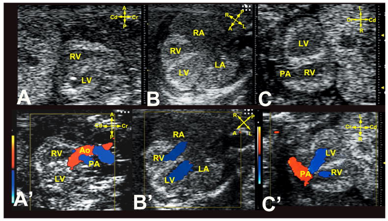
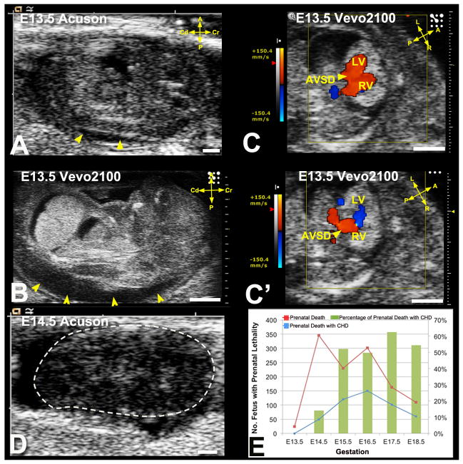
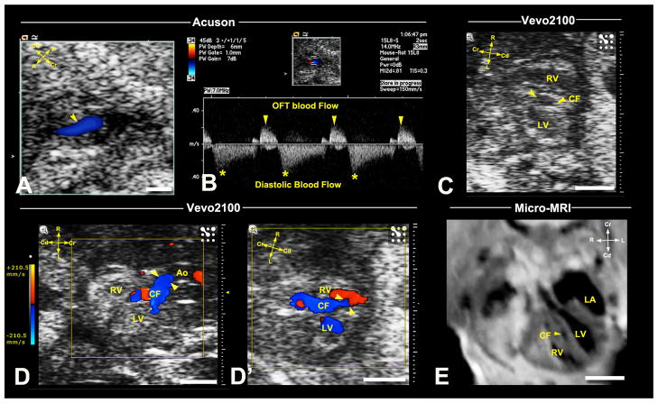
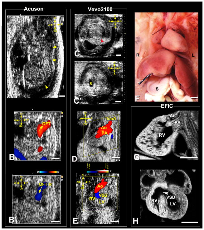
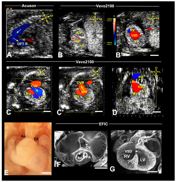
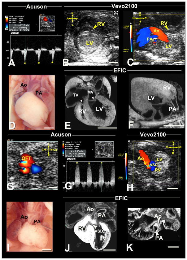
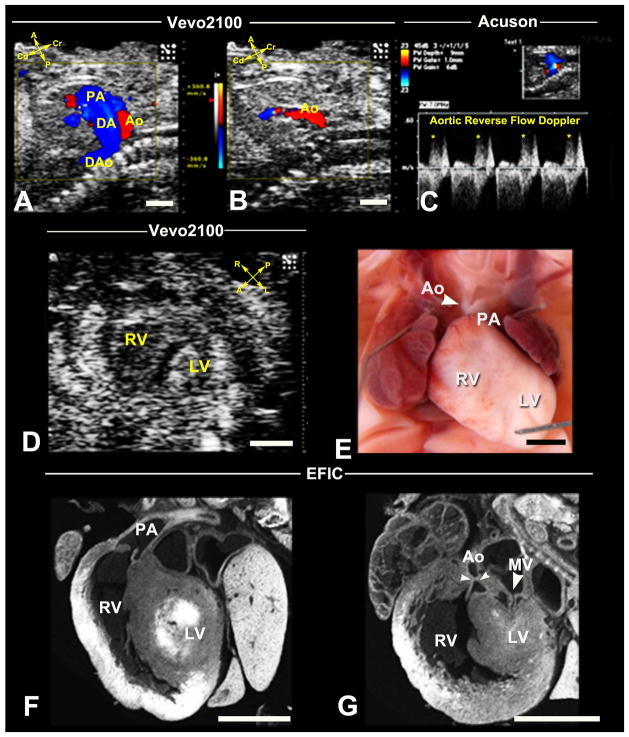
Methods and results: Ethylnitrosourea mutagenized mice were ultrasound-interrogated by fetal echocardiography using a clinical ultrasound system, and fetuses suspected to have cardiac abnormalities were further interrogated with an ultrahigh-frequency ultrasound biomicroscopy. Scanning of 46 270 fetuses revealed 1722 with cardiac anomalies, with 27.9% dying prenatally. Most of the structural heart defects can be diagnosed using ultrasound biomicroscopy but not with the clinical ultrasound system. Confirmation with analysis by necropsy and histopathology showed excellent diagnostic capability of ultrasound biomicroscopy for most CHDs. Ventricular septal defect was the most common CHD observed, whereas outflow tract and atrioventricular septal defects were the most prevalent complex CHD. Cardiac/visceral organ situs defects were observed at surprisingly high incidence. The rarest CHD found was hypoplastic left heart syndrome, a phenotype never seen in mice previously.

Conclusions: We developed a high-throughput, 2-tier ultrasound phenotyping strategy for efficient recovery of even rare CHD phenotypes, including the first mouse models of hypoplastic left heart syndrome. Our findings support a genetic pathogenesis for a wide spectrum of CHDs and suggest that the disruption of left-right patterning may play an important role in CHD.

Keywords: heart defects, congenital; microscopy, acoustic.


Figures

Figure 1
Figure 1. Ultrasound imaging planes for cardiovascular phenotying
Cardiac diagnosis with the UBM is carried out using sagittal (A,A′), transverse four-chamber (B,B′), and frontal (C,C′) imaging planes. Note the anterior-posterior (A–P) axis is equivalent to the body’s dorsal-ventral axis. A, anterior; P, posterior; L, left; R, right; Cr, cranial; Cd, caudal.
Figure 2
Figure 2. Ultrasound detection of prenatal lethality
(A–D) Acuson (A) and Vevo2100 (B) imaging of an E13.5 fetus showed hydrops (A,B, arrowheads). Vevo2100 also showed AVSD (arrowhead in C) with common valve regurgitation (C′ arrowhead). This fetus was found dead the next day (D). (E). Prenatal death with and without CHD detected by ultrasound screening. Scale bar=1mm.
Figure 3
Figure 3. Ultrasound detection of coronary fistula
Acuson imaging detected an E16.5 fetus with abnormal blood flow (arrowhead) associated with the OFT (A), which spectral Doppler showed occurred at diastole (B). Vevo2100 (C,D,D′) imaging in the frontal view revealed a vessel in the ventricular septum (C), which color flow confirmed is a coronary fistula (CF) originating from the aortic root (arrowheads in D), connecting to the RV (arrowheads, D′). This vessel was also visualized by micro-MRI (E,arrowhead). Scale bar=1mm.
Figure 4
Figure 4. Ultrasound diagnosis of heterotaxy with Taussig-Bing DORV
Acuson imaging (A,B,B′) showed an E15.5 fetus with hydrops (arrowheads) and OFT regurgitation (OFT R). Vevo2100 imaging (C,C′) showed heterotaxy with heart apex on left (C,arrowhead) and stomach to right (S,C′), which was confirmed by necropsy (F). Vevo2100 (D,E) and EFIC (G,H) imaging showed aorta (Ao) anterior and PA posterior with both outflows connected to RV (D,G,E), with large VSD (E,H). L, left; R, right; InnA, innominate artery; Scale bar=1mm.
Figure 5
Figure 5. Ultrasound diagnosis of persistent truncus arteriosus
Acuson spectral Doppler showed an E17.5 fetus OFT regurgitation (A,arrowhead; OFT asterisks, forward flow). Vevo2100 revealed a single OFT overriding the ventricular septum with a VSD (arrowhead B). This was associated with OFT regurgitation, indicated by red forward and blue reverse blood flow (B,B′), further confirmed with necropsy (C) and EFIC histology (D,E). A single OFT (CT in C,D) was observed overriding the septum with ventricular noncompaction, a perimembranous (D,arrowhead) and muscular VSDs (arrowheads in D,E). Scale bar=1mm.
Figure 6
Figure 6. Diagnosis of DORV with hypoplastic PA and AVSD
Acuson imaging detected an E16.5 fetus OFT regurgitation (OFT R in A). Vevo2100 imaging (B,B′) revealed a RV-LV shunt, indicating a VSD (arrowheads B). Vevo2100 imaging in the transverse view (C,C′) showed IFT forward (C,arrow) and regurgitant (C′,arrow) flow, while the sagittal view (D) showed majority of the aorta connects to the RV with an adjacent small vessel (arrowhead, PA) and a VSD. Necropsy (E) and EFIC histology (F,G) revealed a hypoplastic PA (E–G) and large VSD (arrowhead,G). A common atrioventricular valve (CV in F) was observed in the transverse view (F). CVF, CVR: common atrioventricular valve forward flow, reverse flow. Scale bar=1mm.
Figure 7
Figure 7. Diagnosis of right and left heart obstructive lesions
(A–F) Hypoplastic right heart syndrome. Acuson Doppler imaging detected an E17.5 fetus with increased velocity in the IFT (A, asterisks). Vevo2100 revealed a hypoplastic RV (B,C) with hypoplastic PA. Necropsy (D) of the stillborn pup confirmed hypoplastic PA, with EFIC imaging (E,F) also showing a hypoplastic RV, hypoplastic tricuspid valve (TV) compared to the mitral valve (MV) (arrowheads,E) and hypoplastic PA connected to RV (arrowhead,F). (G–K) Hypoplastic ascending aorta with VSD. Acuson color flow revealed an E17.5 fetus with OFT aliasing (G), which spectral Doppler (G′) showed high velocity (G′,asterisks). Vevo2100 color flow (H) revealed aortic stenosis with VSD, consistent with the spectral Doppler (G′) findings. Necropsy (I) and EFIC (J,K) histology showed hypoplastic ascending aorta. Also note the VSD (J) and small RV with hypertrophy (J). Scale bar=1mm.
Figure 8
Figure 8. Recovery of HLHS mutant by ultrasound phenotyping
Vevo2100 in sagittal view (A) showed an E16.5 fetus with reversed aortic blood flow. This is seen in diastole (B), and was confirmed by spectral Doppler (C). Vevo2100 imaging in the short axis view (D) showed hypoplastic LV. Necropsy of the stillborn pup (E) revealed hypoplastic aorta and small LV, confirmed by EFIC histology (F,G) to be HLHS with small mitral valve orifice, small LV and hypoplastic aorta. DA, ductus artery; DAo, descending aorta. Scale bar=1mm.

References

    1. Justice MJ. Capitalizing on large-scale mouse mutagenesis screens. Nat Rev Genet. 2000;1:109–115. - PubMed
    1. Shen Y, Leatherbury L, Rosenthal J, Yu Q, Pappas MA, Wessels A, Lucas J, Siegfried B, Chatterjee B, Svenson K, Lo CW. Cardiovascular phenotyping of fetal mice by noninvasive high-frequency ultrasound facilitates recovery of ENU-induced mutations causing congenital cardiac and extracardiac defects. Physiol Genomics. 2005;24:23–36. - PubMed
    1. Yu Q, Shen Y, Chatterjee B, Siegfried BH, Leatherbury L, Rosenthal J, Lucas JF, Wessels A, Spurney CF, Wu YJ, Kirby ML, Svenson K, Lo CW. ENU induced mutations causing congenital cardiovascular anomalies. Development. 2004;131:6211–6223. - PubMed
    1. Rosenthal J, Mangal V, Walker D, Bennett M, Mohun TJ, Lo CW. Rapid high resolution three dimensional reconstruction of embryos with episcopic fluorescence image capture. Birth Defects Res C Embryo Today. 2004;72:213–223. - PubMed
    1. Tobita K, Liu X, Lo CW. Imaging modalities to assess structural birth defects in mutant mouse models. Birth Defects Res C Embryo Today. 2010;90:176–184. - PubMed

Publication types

MeSH terms

Substances