. 2014 Apr 15;23(8):2189-97.
doi: 10.1093/hmg/ddt616. Epub 2013 Dec 6.

A missense mutation in ITGB6 causes pitted hypomineralized amelogenesis imperfecta

Affiliations

A missense mutation in ITGB6 causes pitted hypomineralized amelogenesis imperfecta

James A Poulter et al. Hum Mol Genet. .

Abstract

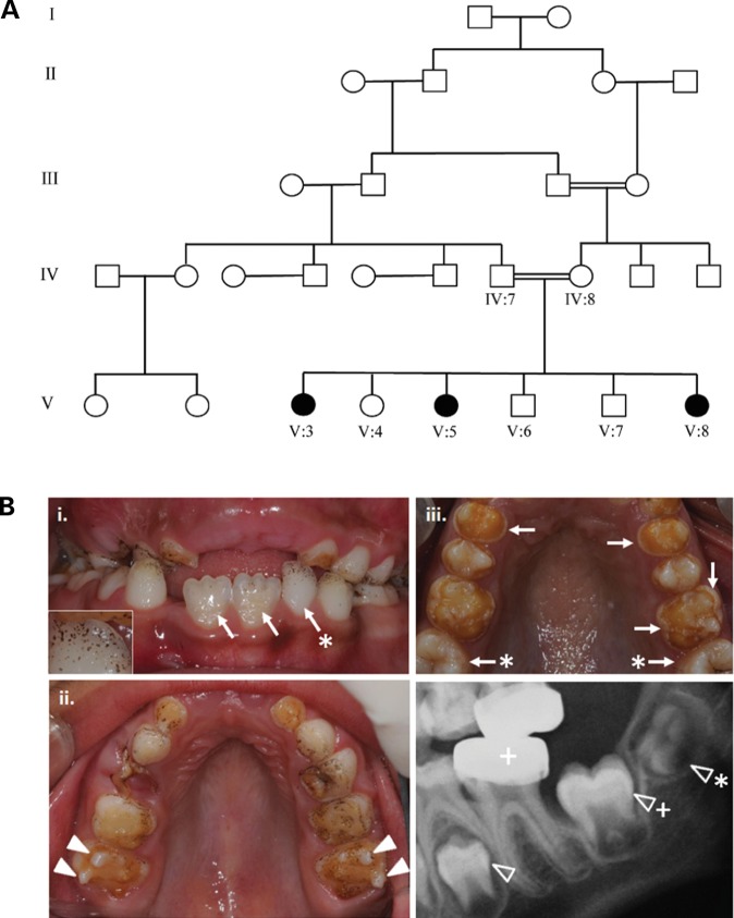
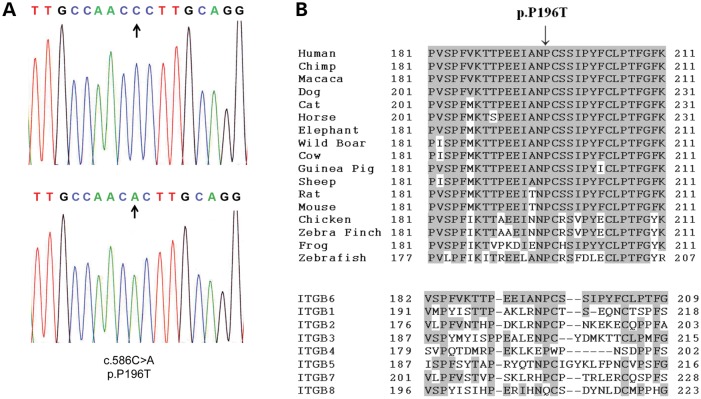
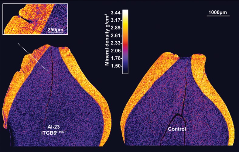
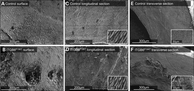
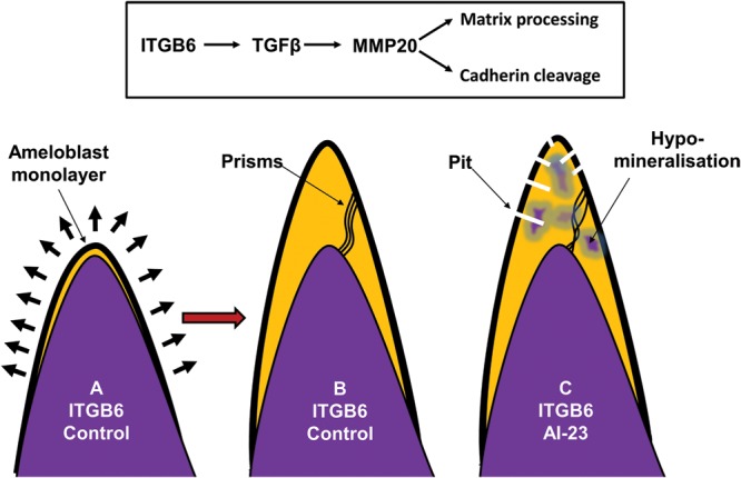
We identified a family in which pitted hypomineralized amelogenesis imperfecta (AI) with premature enamel failure segregated in an autosomal recessive fashion. Whole-exome sequencing revealed a missense mutation (c.586C>A, p.P196T) in the I-domain of integrin-β6 (ITGB6), which is consistently predicted to be pathogenic by all available programmes and is the only variant that segregates with the disease phenotype. Furthermore, a recent study revealed that mice lacking a functional allele of Itgb6 display a hypomaturation AI phenotype. Phenotypic characterization of affected human teeth in this study showed areas of abnormal prismatic organization, areas of low mineral density and severe abnormal surface pitting in the tooth's coronal portion. We suggest that the pathogenesis of this form of AI may be due to ineffective ligand binding of ITGB6 resulting in either compromised cell-matrix interaction or compromised ITGB6 activation of transforming growth factor-β (TGF-β) impacting indirectly on ameloblast-ameloblast interactions and proteolytic processing of extracellular matrix proteins via MMP20. This study adds to the list of genes mutated in AI and further highlights the importance of cell-matrix interactions during enamel formation.


Figures

Figure 1.
Figure 1.
Family pedigree and clinical dental phenotypes for AI-23. (A) Pedigree of family AI-23 recording the three affected individuals within a consanguineous family. DNA was available for all labelled individuals. (B) (i) and (ii) The clinical appearances for V8 aged 7 years of the early mixed dentition with premature enamel failure. Surface enamel pitting is evident on many teeth, including the partially erupted permanent lower incisors (arrows) and via the ‘speckled black’ appearances due to exogenous staining in the pits. Inset image is a higher magnification image of the deciduous tooth (arrow *) highlighting the pitting. The retention of enamel over the cusps of the permanent molar teeth (arrow heads) at this stage highlights the dramatic loss of enamel from the rest of the dental crown, even though these teeth have been in the mouth for a short period of time. (iii) Clinical appearances of the upper dentition for V3 aged 9 years illustrating how the enamel can fracture cleanly away leaving a shoulder of remaining enamel at the cervical margin (arrows). The enamel of the newly erupted second permanent molar teeth has yet to fail (arrow *). (iv) Dental radiograph of V3 aged 7 years confirming a near-normal enamel volume in the unerupted second premolar (arrowhead) and second molar (arrowhead +) lower permanent teeth with a clear difference in radiodensity between enamel and dentine, consistent with what is observed in normal teeth. The first lower permanent molar tooth has been restored with a metal crown (+). The crown of the permanent lower third molar tooth is starting to mineralize (arrowhead *).
Figure 2.
Figure 2.
Electropherogram of mutation and conservation of the P196T variant. (A) Representative electropherogram of the ITGB6 mutation in an affected member of family AI-23 alongside the wild-type sequence. Arrows indicate the localization of the variant. (B) Conservation of the P196 residue in orthologues (upper) and paralogues (lower). Conserved residues are highlighted. Human (NP_000879), Chimp (XP_001149234), Macaca (XP_001094740), Dog (XP_857148), Cat (XP_003990848), Horse (XP_001492914), African Elephant (XP_003406050), Wild Boar (NP_001090892), Cow (NP_777123), Guinea Pig (XP_003478725), Sheep (NP_001107244), Rat (NP_001004263), Mouse (NP_067334), Chicken (XP_422037), Zebra Finch (XP_002193421), Frog (NP_001090775), Zebrafish (XP_003199474), Human ITGB1 (NP_596867), ITGB2 (NP_001120963), ITGB3 (NP_000203), ITGB4 (NP_000204), ITGB5 (NP_002204), ITGB7 (NP_000880) and ITGB8 (NP_002205).
Figure 3.
Figure 3.
Phenotypic analyses of enamel: µCT. (A) µCT confirmed a reasonable enamel volume in affected teeth, but with a multiple enamel surface and sub-surface abnormalities. Particularly striking were the regions of enamel exhibiting reduced mineral density and pits running for the enamel surface deep in to the bulk of the enamel.
Figure 4.
Figure 4.
Phenotypic analyses of enamel: SEM. (A) SEM of the etched control enamel surface showed the characteristic appearance of arrays of enamel prisms terminating at the surface. (B) In contrast, the affected enamel was punctured by numerous pits and the prism array was more obscure. (C) SEM of the internal prim architecture of control enamel in longitudinal section revealed the characteristic sinusoidal pattern of prism cohorts giving rise to Hunter–Schreger bands. (D) The prism architecture in affected enamel was less regular and clear Hunter–Schreger bands were less distinct. The inset images in (C) and (D) show that the individual prism structure at the micron level in control and affected enamel is indistinguishable by SEM in some areas. (E) SEM of transversely cut sections through control cuspal enamel showed the characteristic array of prisms themselves in the transverse section (higher magnification inset). (F) In affected enamel, the inner enamel layer is structurally abnormal with loss of prism organization. Structural features designed to provide mechanical stability that depend on the correct inter-relationship between prism cohorts (e.g. Hunter–Schreger bands) will be compromised in this enamel (inset shows higher magnification details).
Figure 5.
Figure 5.
Cartoon summarizing the hypothesis presented here to explain the mechanism underpinning the AI subtype described. (A) At the beginning of normal enamel secretion, an army of ameloblasts, present as a monolayer, migrates away from the preformed dentine surface leaving the enamel matix in their wake. (B) As the cuspal enamel volume increases the ameloblasts modulate cell–cell contacts to cope with the stresses encountered by the monolayer being required to cover an ever expanding area. We hypothesize that ITGB6 upregulates MMP20 expression (via TGFβ activation). This in turn cleaves cadherin, thus allowing ameloblasts to modulate cell–cell contacts to cope with the increasing stress of an expanding enamel surface and to allow cohorts of ameloblast to move relative to one another to produce a sinusoidal prism architecture. MMP20 also processes enamel matrix proteins, which is required for their final degradation prior to the completion of mineralization. In affected enamel, we hypothesize that cadherin cleavage and matrix processing are compromised due to the ITGB6 mutation resulting in breaks in the ameloblast monolayer in the cuspal regions leading to pitting, abnormal prism architecture and retained matrix proteins that inhibit final enamel mineralization.

References

    1. Bailleul-Forestier I., Molla M., Verloes A., Berdal A. The genetic basis of inherited anomalies of the teeth. Part 1: clinical and molecular aspects of non-syndromic dental disorders. Eur. J. Med. Genet. 2008;51:273–291. - PubMed
    1. Coffield K.D., Phillips C., Brady M., Roberts M.W., Strauss R.P., Wright J.T. The psychosocial impact of developmental dental defects in people with hereditary amelogenesis imperfecta. J. Am. Dent. Assoc. 2005;136:620–630. - PubMed
    1. Robinson C., Kirkham J., Weatherell J.A., Richards A., Josephsen K., Fejerskov O. Mineral and protein concentrations in enamel of the developing permanent porcine dentition. Caries Res. 1988;22:321–326. - PubMed
    1. Smith C.E. Cellular and chemical events during enamel maturation. Crit. Rev. Oral Biol. Med. 1998;9:128–161. - PubMed
    1. Sawada T., Yamamoto T., Yanagisawa T., Takuma S., Hasegawa H., Watanabe K. Evidence for uptake of basement membrane by differentiating ameloblasts in the rat incisor enamel organ. J. Dent. Res. 1990;69:1508–1511. - PubMed

Publication types

MeSH terms