. 2014 Mar;85(3):472-81.
doi: 10.1124/mol.113.089516. Epub 2013 Dec 6.

Regulation of β2-adrenergic receptor function by conformationally selective single-domain intrabodies

Affiliations

Regulation of β2-adrenergic receptor function by conformationally selective single-domain intrabodies

Dean P Staus et al. Mol Pharmacol. 2014 Mar.

Abstract

The biologic activity induced by ligand binding to orthosteric or allosteric sites on a G protein-coupled receptor (GPCR) is mediated by stabilization of specific receptor conformations. In the case of the β2 adrenergic receptor, these ligands are generally small-molecule agonists or antagonists. However, a monomeric single-domain antibody (nanobody) from the Camelid family was recently found to allosterically bind and stabilize an active conformation of the β2-adrenergic receptor (β2AR). Here, we set out to study the functional interaction of 18 related nanobodies with the β2AR to investigate their roles as novel tools for studying GPCR biology. Our studies revealed several sequence-related nanobody families with preferences for active (agonist-occupied) or inactive (antagonist-occupied) receptors. Flow cytometry analysis indicates that all nanobodies bind to epitopes displayed on the intracellular receptor surface; therefore, we transiently expressed them intracellularly as "intrabodies" to test their effects on β2AR-dependent signaling. Conformational specificity was preserved after intrabody conversion as demonstrated by the ability for the intracellularly expressed nanobodies to selectively bind agonist- or antagonist-occupied receptors. When expressed as intrabodies, they inhibited G protein activation (cyclic AMP accumulation), G protein-coupled receptor kinase (GRK)-mediated receptor phosphorylation, β-arrestin recruitment, and receptor internalization to varying extents. These functional effects were likely due to either steric blockade of downstream effector (Gs, β-arrestin, GRK) interactions or stabilization of specific receptor conformations which do not support effector coupling. Together, these findings strongly implicate nanobody-derived intrabodies as novel tools to study GPCR biology.


Figures

Fig. 1.
Fig. 1.
Identification of conformationally selective β2AR nanobodies. (A) Classification of β2AR nanobodies based on CDR3 conservation. Nanobodies with a divergent CDR3 were categorized as MISC. (B) ELISA assay describing nanobody selectivity for active agonist-bound (BI-167107) or inactive antagonist-bound (ICI-118551) β2AR. Using immobilized nanobody, the relative capture of BI-167107–β2AR was subtracted from that derived with ICI-118551–β2AR; values greater than 0 (blue bars) denote nanobodies that preferentially bound BI-167107–β2AR, whereas negative values represent preference for ICI-118551–β2AR. The absence of nanobody (—) and Nb30 were negative controls. T-tests were performed to determine the significance between each nanobody and (—) (*P < 0.05).
Fig. 2.
Fig. 2.
Nanobodies allosterically stabilize active or inactive β2AR conformations. Sf9 insect cell membranes expressing FLAG-β2AR were incubated with 4 nM [3H]methoxyfenoterol and 1 µM Nb in the absence (total binding) or presence of 10 µM propranolol (nonspecific). Specific binding was determined by subtracting nonspecific from total binding and normalized relative to Nb80. The absence of nanobody (—) and Nb30 were negative controls. t tests were performed to determine the significance between each nanobody and (—) (*P < 0.05).
Fig. 3.
Fig. 3.
Nanobodies bind specifically to intracellular epitopes of the β2AR. Sf9 insect cells were infected with a baculovirus encoding FLAG-β2AR resulting in cell populations that were partially permeabilized due to viral infection. β2AR-expressing cells were preincubated with 1 µM ICI-118551 (family A nanobodies) or 1 µM BI-167107 (families B, C, and MISC). Binding of purified His-tagged nanobodies to cells was detected with a DyLight488-labeled anti–6× His antibody. FLAG-M1 antibody was labeled with DyLight488. Singlet cells were gated into intact and permeable populations based on staining with SYTOX AADvanced Dead Cell Stain.
Fig. 4.
Fig. 4.
Intrabody 80 expression and specificity for activated β2AR. (A) Immunoblotting (IB) of HEK293 cells transiently transfected with FLAG-β2AR and pcDNA, HA-Ib30, or Ib80. (B) HEK293 cells were transfected as described earlier, and receptor expression was assessed via whole-cell binding using [3H]CGP-12177A. Specific binding normalized to pcDNA. (C) HEK293 cells stably expressing FLAG-β2AR were transfected with HA-Ib30 or Ib80, stimulated with the agonist ISO for 15 minutes, and then solubilized in lysis buffer. FLAG-β2AR was immunoprecipitated (IP) using FLAG beads, the eluate was subjected to SDS-PAGE, and intrabody was detected using an HA antibody.
Fig. 5.
Fig. 5.
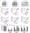
Regulation of β2AR functions by “active” state stabilizing intrabodies. Receptor expression (A), G protein–mediated cAMP levels (GloSensor) (B), and β-arrestin recruitment (Tango Assay) (C) was measured for intrabody family B, C, and MISC. (A) Immunoblot analysis of HEK293 cells transiently transfected with FLAG-β2AR and the indicated HA-Intrabody (Ib). Tubulin was used to ensure equal total protein loading. (B) HEK293 cells were transfected with the GloSensor cAMP biosensor (Promega) and pcDNA empty vector or individual HA-intrabodies, stimulated with a dose response of isoproterenol (ISO), and luminescence measured 10 minutes thereafter. Data were analyzed using GraphPad Prism program with sigmoidal dose response curve fit and normalized to 100% pcDNA empty vector. (C) HEK293T cells stably expressing β-arrestin-2 fused to the Tobacco Etch Virus (TEV) protease and a tTA transcription factor driven luciferase reporter were transiently transfected with β2AR fused to the tTA transcription factor, but separated by a TEV cleavage site and the indicated HA-intrabody. Cells were stimulated with a dose response of isoproterenol, and luminescence was measured 16 hours thereafter. Data were normalized as described in C. (D) The maximal response (Emax) for cAMP and β-arrestin following isoproterenol treatment in presence of the indicated intrabody as determined by nonlinear regression analysis. Data were normalized to 100% pcDNA. T-tests were used to compare each intrabody to pcDNA control (*P < 0.05), underlined • denotes significance between Bmax of Glosensor and Tango assays for each individual intrabody
Fig. 6.
Fig. 6.
Intrabodies that stabilize “inactive” β2AR conformation(s) inhibit G protein activation and β-arrestin recruitment. The effects of intrabody family A on expression of β2AR (A), G protein activation (B), and β-arrestin recruitment (C) were analyzed as described in Fig. 4. (D) The maximal response (Emax) for cAMP and β-arrestin following isoproterenol treatment in the presence of indicated intrabody as determined by nonlinear regression analysis. Data were normalized to 100% pcDNA. t tests were used to compare each intrabody to pcDNA control (*P < 0.05); underlined • denotes significance between Bmax of GloSensor and Tango assays for each individual intrabody.
Fig. 7.
Fig. 7.
Attenuation of β-arrestin recruitment, GRK-mediated receptor phosphorylation, and β2AR internalization by intrabody 71. (A) HEK293 cells were transfected with FLAG-β2AR and pcDNA empty vector (EV) or HA-Ib71, and cell surface expression was measured via [3H]CGP-12177A radioligand binding. Data were normalized relative to pcDNA. (B) HA-Ib71 was transfected in HEK293 cells stably expressing FLAG-β2AR, and coimmunoprecipitation assays were performed after treatment with DMSO (−) or ISO (+). (C) HEK293 cells expressing FLAG-β2AR and pcDNA or HA-Ib71 were subject to whole-cell binding experiments in the presence of [3H]CGP-12177A and a dose response of isoproterenol. (D) HEK293 cells transiently expressing FLAG-β2AR and pcDNA, Ib30, or Ib71 were stimulated with a dose response of isoproterenol and analyzed for GRK-dependent phosphorylation of serine 355/6. (E) Whole-cell cross-linking of β-arrestin-1/2 with FLAG-β2AR in HEK293 cells in the presence of pcDNA, Ib30, or Ib71 after stimulation with 10 µM isoproterenol. (F) U2OS cells were transiently transfected with FLAG-β2AR, green fluorescent protein–β-arrestin-2, and Myc-tagged Ib30 or Ib71, and β-arrestin recruitment and receptor internalization were visualized using immunostaining and confocal microscopy. DAPI, 4′,6-diamidino-2-phenylindole; IB, immunoblots; IP, immunoprecipitation.

References

    1. Barlow JN, Conrath K, Steyaert J. (2009) Substrate-dependent modulation of enzyme activity by allosteric effector antibodies. Biochim Biophys Acta 1794:1259–1268 - PubMed
    1. Barnea G, Strapps W, Herrada G, Berman Y, Ong J, Kloss B, Axel R, Lee KJ. (2008) The genetic design of signaling cascades to record receptor activation. Proc Natl Acad Sci USA 105:64–69 - PMC - PubMed
    1. Benovic JL, Kühn H, Weyand I, Codina J, Caron MG, Lefkowitz RJ. (1987) Functional desensitization of the isolated beta-adrenergic receptor by the beta-adrenergic receptor kinase: potential role of an analog of the retinal protein arrestin (48-kDa protein). Proc Natl Acad Sci USA 84:8879–8882 - PMC - PubMed
    1. Bertin B, Freissmuth M, Jockers R, Strosberg AD, Marullo S. (1994) Cellular signaling by an agonist-activated receptor/Gs alpha fusion protein. Proc Natl Acad Sci USA 91:8827–8831 - PMC - PubMed
    1. Conklin BR, Hsiao EC, Claeysen S, Dumuis A, Srinivasan S, Forsayeth JR, Guettier JM, Chang WC, Pei Y, McCarthy KD, et al. (2008) Engineering GPCR signaling pathways with RASSLs. Nat Methods 5:673–678 - PMC - PubMed

Publication types

MeSH terms

Substances