. 2014 Feb;42(4):2538-54.
doi: 10.1093/nar/gkt1256. Epub 2013 Dec 6.

A cypovirus VP5 displays the RNA chaperone-like activity that destabilizes RNA helices and accelerates strand annealing

Affiliations

A cypovirus VP5 displays the RNA chaperone-like activity that destabilizes RNA helices and accelerates strand annealing

Jie Yang et al. Nucleic Acids Res. 2014 Feb.

Abstract

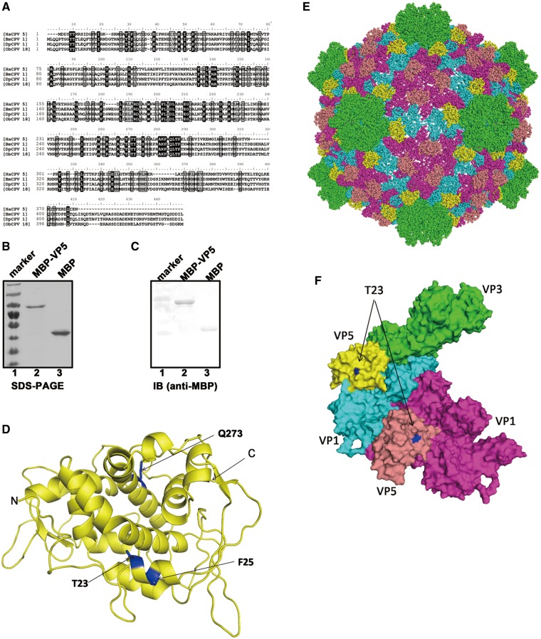
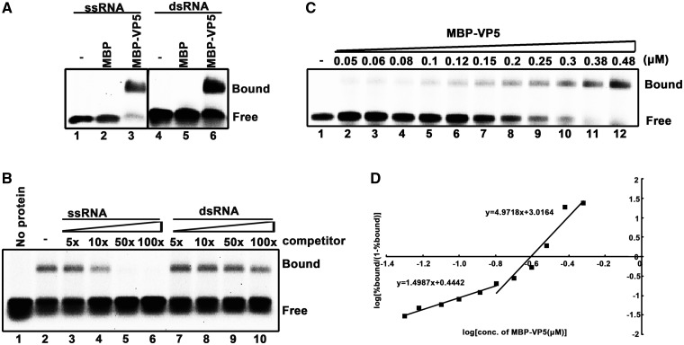
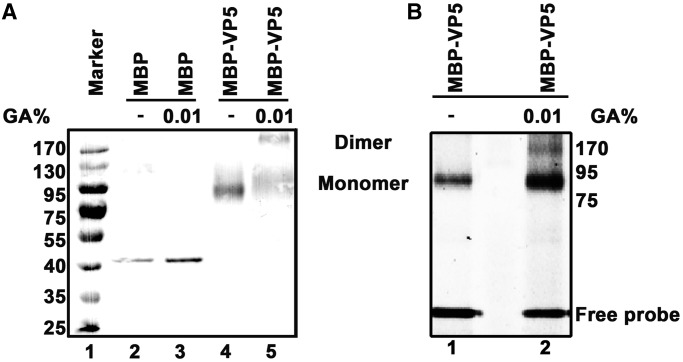
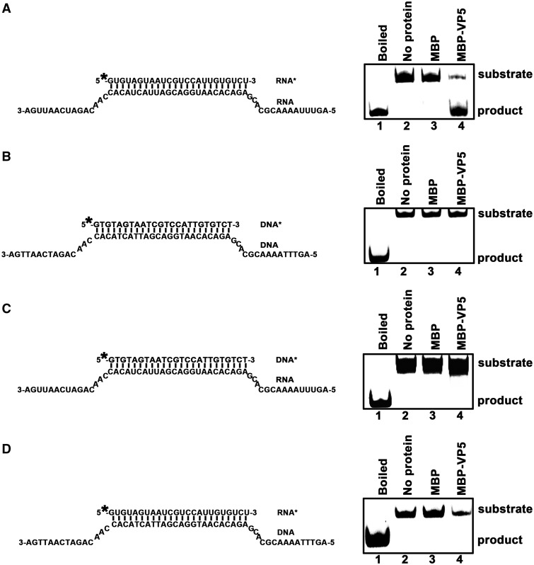
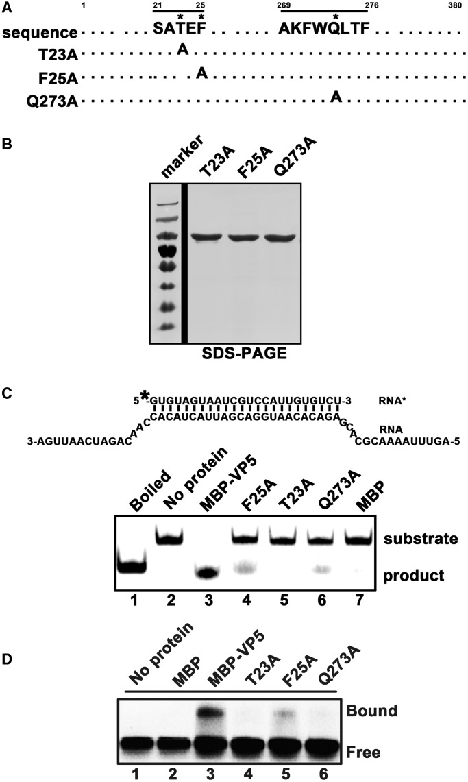
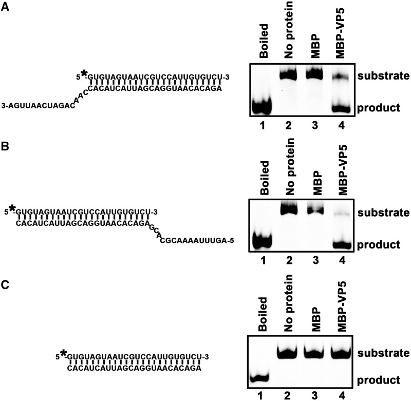
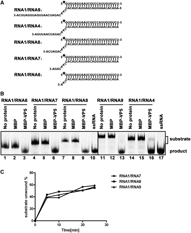
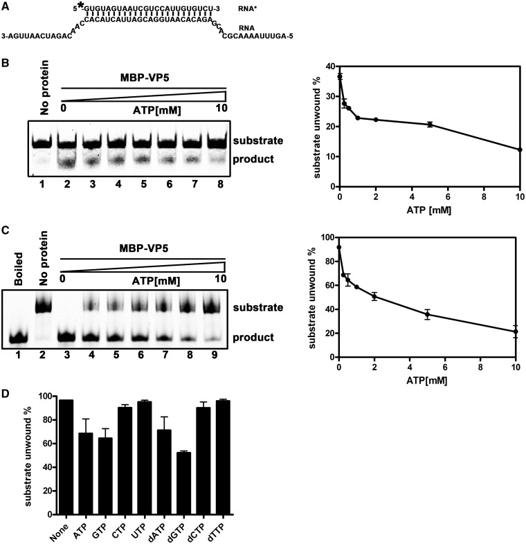
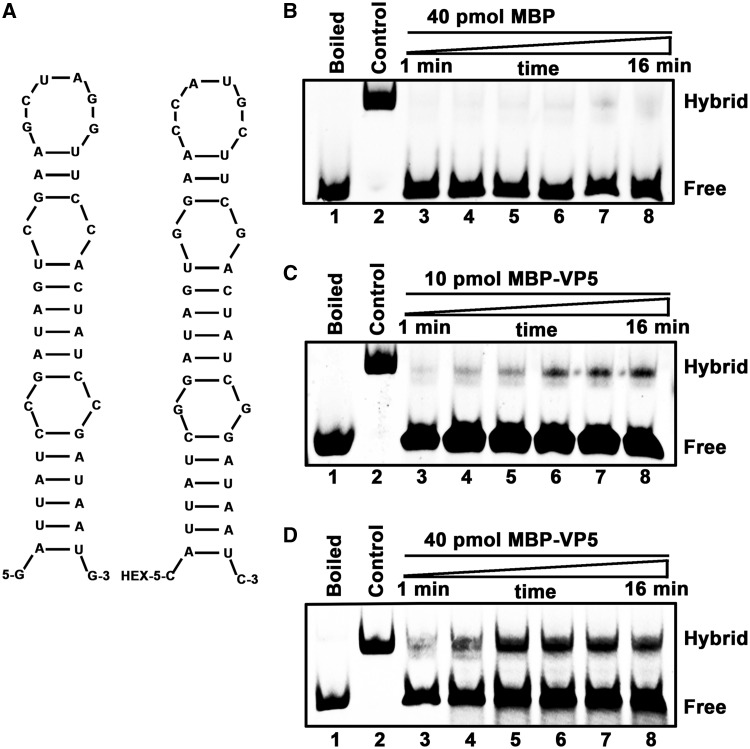
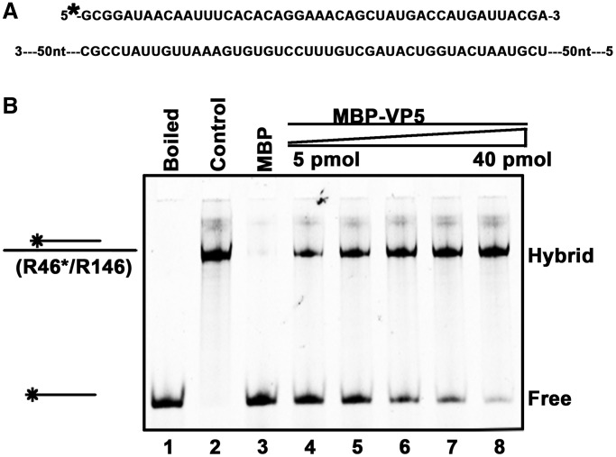
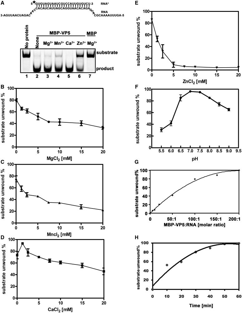
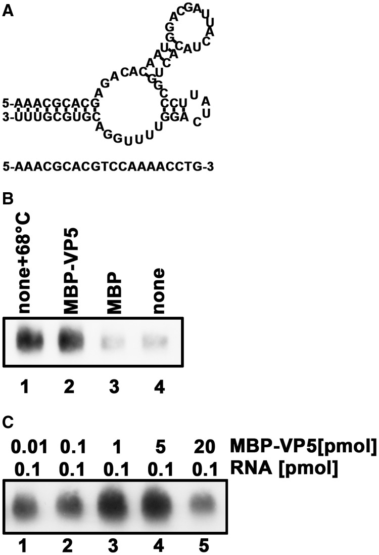
For double-stranded RNA (dsRNA) viruses in the family Reoviridae, their inner capsids function as the machinery for viral RNA (vRNA) replication. Unlike other multishelled reoviruses, cypovirus has a single-layered capsid, thereby representing a simplified model for studying vRNA replication of reoviruses. VP5 is one of the three major cypovirus capsid proteins and functions as a clamp protein to stabilize cypovirus capsid. Here, we expressed VP5 from type 5 Helicoverpa armigera cypovirus (HaCPV-5) in a eukaryotic system and determined that this VP5 possesses RNA chaperone-like activity, which destabilizes RNA helices and accelerates strand annealing independent of ATP. Our further characterization of VP5 revealed that its helix-destabilizing activity is RNA specific, lacks directionality and could be inhibited by divalent ions, such as Mg(2+), Mn(2+), Ca(2+) or Zn(2+), to varying degrees. Furthermore, we found that HaCPV-5 VP5 facilitates the replication initiation of an alternative polymerase (i.e. reverse transcriptase) through a panhandle-structured RNA template, which mimics the 5'-3' cyclization of cypoviral positive-stranded RNA. Given that the replication of negative-stranded vRNA on the positive-stranded vRNA template necessitates the dissociation of the 5'-3' panhandle, the RNA chaperone activity of VP5 may play a direct role in the initiation of reoviral dsRNA synthesis.


Figures

Figure 1.
Figure 1.
(A) The amino acid sequence alignment of VP5 proteins of HaCPV-5, BmCPV-1, Dendrolimus punctatus CPV-1 (DpCPV-1) and Operophtera brumata CPV-18 (ObCPV-18). Multiple sequence alignments were generated using ClustalX. (B) Electrophoresis analysis of purified MBP-VP5 and MBP alone. Proteins were loaded onto a 12% SDS-PAGE gel and then visualized via Coomassie blue staining. Lane 1, protein molecular mass markers; lane 2, purified MBP-VP5; lane 3, purified MBP. (C) The purified MBP-VP5 and MBP alone were subjected to SDS-PAGE, followed by western blot analysis with anti-MBP polyclonal antibodies. (D) The 3D structure of HaCPV-5 VP5 was modeled by the HMMSTR/Rosetta server, and drawn by PyMOL as described in ‘Materials and Methods’ section. The three mutation sites as well as N- and C-termini were labeled as indicated. (E and F) The surface representations of modeled VP5, which indicate the orientation of VP5 on the CPV particle (E), or in the asymmetric unit formed by VP1, VP3 and VP5 (F). The T23 on the VP5 is shown in blue. Color-coded by protein subunits, VP3 is in yellow, VP1 has two conformers in cyan and magenta, while VP5 in yellow and salmon. The maps of CPV particle and the asymmetric unit are drawn by PyMOL based on the atomic cryoEM structure of BmCPV capsid (PDB number 3IZX) by replacing BmCPV VP5 with HaCPV-5 VP5.
Figure 2.
Figure 2.
VP5 binds to RNA in a cooperative manner. (A) A gel mobility shift assay was performed to evaluate the dsRNA or ssRNA binding capacity of MBP-VP5. Ten picomoles MBP-VP5 was incubated with 0.1 pmol HEX-labeled ssRNA (RNA1) or dsRNA (RNA1/RNA2) substrate for 30 min. Lanes 1 and 4, no protein supplemented; lanes 2 and 5, 10 pmol MBP supplemented; lanes 3 and 6, 10 pmol MBP-VP5 supplemented. Protein-bound and free RNA strands are indicated. (B) 0.1 pmol HEX-labeled ssRNA probe and 2 pmol MBP-VP5 were incubated with unlabeled competitor ssRNA or dsRNA at the indicated increasing concentrations (in 5-, 10-, 50-, 100-folded excess over the amount of HEX-labeled ssRNA). The bound complexes were analyzed in a gel mobility shift assay. (C) Gel mobility shift assays were performed by incubating the indicated increasing concentrations (0.05–0.45 μM) of MBP-VP5 with 0.1 pmol RNA1 probe. (D) The RNA binding data in (C) were quantified, and a Hill transformation was applied. The Hill coefficients of the RNA binding of VP5 at low and high protein concentrations are indicated.
Figure 3.
Figure 3.
Monomeric and dimeric MBP-VP5 bind to ssRNA. (A) MBP alone (lane 3) or MBP-VP5 (lane 5) was incubated in a chemical cross-linking buffer containing 0.01% glutaraldehyde (GA) as indicated, and then analyzed via 10% SDS-PAGE, followed by western blots with anti-MBP antibodies as described in ‘Materials and Methods’ section. Lanes 2 and 4, MBP alone and MBP-VP5 in the absence of 0.01% GA. (B) MBP-VP5 was firstly UV cross-linked with HEX-labeled RNA1 probe, and then subjected to chemical cross-linking by incubating with 0.01% GA. The samples were analyzed via 10% SDS-PAGE, followed by scanning using a Typhoon 9200 imager to visualize HEX-labeled RNA probe. The dimeric and monomeric forms of MBP-VP5 are indicated.
Figure 4.
Figure 4.
VP5 destabilizes RNA helices. Purified MBP-VP5 was incubated with (A) standard RNA helix (R*/R substrate), (B) DNA helix (D*/D substrate), (C) DNA/RNA hybrid helix (D*/R substrate) or (D) RNA/DNA hybrid helix (R*/D substrate) as illustrated in the left panels. Asterisk indicates HEX-labeled strands. The preparations of these destabilizing substrates are indicated in ‘Materials and Methods’ section. Helix substrate (0.1 pmol) was incubated in standard reaction mixtures in the presence or absence of 10 pmol MBP-VP5 as indicated, and the destabilizing activity was determined via gel electrophoresis and scanning on a Typhoon 9200. Lane 1, boiled reaction mixture without protein supplementation; lane 2, reaction mixture without protein supplementation; lane 3, reaction mixture with MBP alone; and lane 4, reaction mixture with MBP-VP5.
Figure 5.
Figure 5.
Mutational analysis of the RNA helix-destabilizing activity of VP5. (A) Illustration of the mutagenesis strategy. Asterisk indicates sites of replacement with alanine. (B) Expressed and purified MBP-VP5 mutants were subjected to 12% SDS-PAGE followed by Coomassie brilliant blue R250 staining. (C) Helix substrate (0.1 pmol; RNA1/RNA3; upper panel) was reacted with 10 pmol MBP-VP5 wild-type and mutants as indicated (lanes 3–6). Boiled reaction mixture (lane 1) was used as positive control, and the reaction mixture with no protein (lane 2) or supplemented with MBP alone (lane 7) was used as a negative control. (D) Gel mobility shift assays were performed by incubating 0.1 pmol HEX-labeled RNA1 with 10 pmol MBP-VP5 wild type and mutants as indicated (lanes 3–6). Reaction mixture without protein supplementation (lane 1) or with MBP alone (lane 2) was used as a negative control.
Figure 6.
Figure 6.
VP5 destabilizes RNA helices in a bidirectional manner. Purified MBP-VP5 was incubated with (A) 3′-tailed RNA helix (RNA1/RNA4), (B) 5′-tailed RNA helix (RNA1/RNA5) or (C) blunt-ended RNA helix (RNA1/RNA2) as illustrated in the left panels. Asterisk indicates the HEX-labeled strand (RNA1). The preparations of these destabilizing substrates are indicated in ‘Materials and Methods’ section. RNA helix substrate (0.1 pmol) was incubated in standard reaction mixtures in the presence or absence of 10 pmol MBP-VP5 as indicated, and the destabilizing activity was determined via gel electrophoresis and scanning on a Typhoon 9200. Lane 1, boiled reaction mixture without protein supplementation; lane 2, reaction mixture without protein supplementation; lane 3, reaction mixture with MBP alone; and lane 4, reaction mixture with MBP-VP5.
Figure 7.
Figure 7.
The length of 3′-tail of RNA helices has no obvious impact on the helix-destabilizing activity of VP5. (A) Schematic illustration of RNA helix substrates with the indicated lengths of 3′-tails. The shorter strand (RNA1) was HEX labeled. (B) Indicated 3′-tailed RNA helix substrate (0.1 pmol) was incubated with 10 pmol MBP-VP5, and the destabilizing activity was determined via gel electrophoresis and scanning on a Typhoon 9200. Reaction mixture without protein supplementation (lanes 1, 4, 7, 11 and 14) or with MBP alone (lanes 2, 5, 8, 12 and 15) was used as a negative control, and ssRNA was loaded to indicate the position of free ssRNA strand in the gel (lanes 10 and 17). (C) Three different RNA helix substrates (RNA1/RNA7, RNA1/RNA8 and RNA1/RNA9) were incubated with 10 pmol MBP-VP5 for 0, 5, 10, 20 and 25 min. The unwinding activity was quantified and plotted as the percentage of the released RNA from the total RNA helix substrate (Y-axis) at each time point (X-axis).
Figure 8.
Figure 8.
Increasing concentrations of ATP inhibited the helix-destabilizing activity of VP5. (A) The standard RNA helix substrate (RNA1/RNA3) is indicated, and asterisk indicates the HEX-labeled strand. (B) The helix substrate (0.1 pmol) was incubated with 10 pmol MBP-VP5 for 10 min in the standard reaction condition in the absence (lane 2) or presence of increasing concentrations of ATP (lanes 3–8; 0.25, 0.5, 1.0, 2.0, 5.0 and 10.0 mM ATP). (C) The helix substrate (0.1 pmol) was incubated with 10 pmol MBP-VP5 for 60 min in the standard reaction condition in the absence (lane 3) or presence of increasing concentrations of ATP (lanes 4–9; 0.25, 0.5, 1.0, 2.0, 5.0 and 10.0 mM ATP). The destabilizing activity was determined via gel electrophoresis and scanning on a Typhoon 9200. The unwinding activity was plotted as the percentage of the released RNA from the total RNA helix substrate (Y-axis) at each ATP concentration (X-axis) (B and C, right panels). Error bars represent standard deviation (SD) values from three separate experiments. (D) The helix substrate (0.1 pmol) was incubated with 10 pmol MBP-VP5 for 60 min in the standard reaction condition in the presence of different NTP or dNTP at 0.5 mM as indicated. The unwinding activity was graphed as the percentage of the released RNA from the total substrate (Y-axis) in the presence of different NTP or dNTP (X-axis). Error bars represent SD values from three separate experiments.
Figure 9.
Figure 9.
VP5 destabilizes stem-loop structured RNA strands. (A) Schematic illustrations of the stem-loop structures of the two complementary 42-nt RNA substrates. HEX labeling is indicated (right). (B–D) The two strands were mixed (0.1 pmol each strand) and reacted in the presence of (B) 40 pmol MBP, (C) 10 pmol MBP-VP5 or (D) 40 pmol MBP-VP5 for the indicated time (lanes 3–8; 1, 2, 4, 8, 10 and 16 min). For (B–D), the mix of the two strands was boiled (lane 1) or preannealed (lane 2) as a negative or positive control. The hybridized and free strands are indicated.
Figure 10.
Figure 10.
VP5 stimulates RNA strand annealing. (A) Schematic illustrations of the HEX-labeled 46-nt RNA strand and the nonlabeled longer complementary 146-nt RNA strand. Asterisk indicates the HEX-labeled strand. (B) Equal amounts (0.1 pmol each) of the two strands were mixed and reacted with increasing amounts of MBP-VP5 for 15 min. Lane 1, the mixture was boiled before being loaded onto the gel as a negative control; lane 2, the two strands were preannealed using a thermal cycler as a positive control; lane 3, the reaction with MBP alone as a negative control; and lanes 4–8, the reactions with 5, 10, 15, 20 and 40 pmol MBP-VP5.
Figure 11.
Figure 11.
Optimal biochemical reaction conditions for the RNA helix-destabilizing activity of VP5. (A) Standard RNA helix substrate (RNA1/RNA3; 0.1 pmol) as illustrated in upper panel was incubated with 10 pmol MBP-VP5 in the absence (lane 2) or presence of 2.5 mM indicated divalent metal ions (lanes 3–6) for 60 min. Reaction mixture without protein addition (lane 1) or with MBP alone (lane 7) was used as a negative control. Asterisk indicates the HEX-labeled strand. (B–E) Standard RNA helix substrate (RNA1/RNA3; 0.1 pmol) was incubated with 10 pmol MBP-VP5 in the presence of increasing concentrations of (B) MgCl2, (C) MnCl2, (D) CaCl2 or (E) ZnCl2 as indicated for 30 min. (F) Standard RNA helix substrate (0.1 pmol) was incubated with 10 pmol MBP-VP5 at indicated pH values in the absence of divalent ions for 30 min. (G) Standard RNA helix substrate (0.1 pmol) was incubated with MBP-VP5 at indicated VP5/RNA molar ratios for 30 min. (H) 10 pmol MBP-VP5 was incubated with 0.1 pmol standard RNA helix substrate (at the molar ratio of 100:1) for indicated time intervals. For (B–H), the unwinding activity was plotted as the percentage of the released RNA from the total RNA helix substrate as Y-axis at indicated ion concentrations (B–E), pH values (F), VP5/RNA molar ratios (G) or incubation time intervals (H) as X-axis. Error bars represent SD values from three separate experiments.
Figure 12.
Figure 12.
VP5 facilitates RT initiation via a CPV panhandle. (A) Schematic illustrations of the predicted panhandle structure formed by 36 bases from the 5′-end and 30 bases from the 3′-end of HaCPV-5 RNA segment 8 (upper panel) and the primer complementary to the 3′-end of the panhandle (lower panel). (B) The RT reactions were conducted using the RNA panhandle structure and the DNA primer in the presence of M-MLV reverse transcriptase for 30 min at 25°C in the absence (lanes 1 and 4) or presence of MBP-VP5 (lane 2) or MBP alone (lane 3). For lane 1, the panhandle and primer were prehybridized via a thermal annealing treatment at 68°C before the reaction. (C) The RT reactions were conducted using the indicated amounts of RNA panhandles in the presence of indicated amounts of MBP-VP5 for 30 min at 25°C. The samples were analyzed on 6% urea PAGE, followed by northern blotting.

References

    1. Mertens P. The dsRNA viruses. Virus Res. 2004;101:3–13. - PubMed
    1. Reinisch KM. The dsRNA Viridae and their catalytic capsids. Nat. Struct. Biol. 2002;9:714–716. - PubMed
    1. Xia Q, Jakana J, Zhang JQ, Zhou ZH. Structural comparisons of empty and full cytoplasmic polyhedrosis virus. Protein-RNA interactions and implications for endogenous RNA transcription mechanism. J. Biol. Chem. 2003;278:1094–1100. - PubMed
    1. Poranen MM, Bamford DH. In: Viral Molecular Machines. Rossmann MG, Rao VB, editors. Springer, New York, NY, USA: Science; 2012. pp. 379–402.
    1. King AMQ, Adams MJ, Carstens EB, Lefkowitz EJ. Virus taxonomy: ninth report of the International Committee on Taxonomy of Viruses. Vol. 9. Academic Press, Salt Lake City, UT, USA: Elsevier; 2011. pp. 497–650.

Publication types