. 2013;10(4):247-251.
doi: 10.1007/s10388-013-0382-8. Epub 2013 Jul 13.

A case report of pulmonary tumor thrombotic microangiopathy (PTTM) caused by esophageal squamous cell carcinoma

Affiliations

A case report of pulmonary tumor thrombotic microangiopathy (PTTM) caused by esophageal squamous cell carcinoma

Takeshi Fujishiro et al. Esophagus. 2013.

Abstract

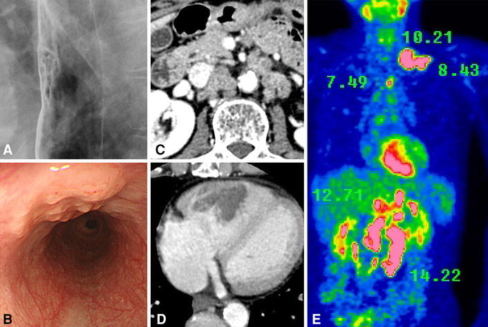
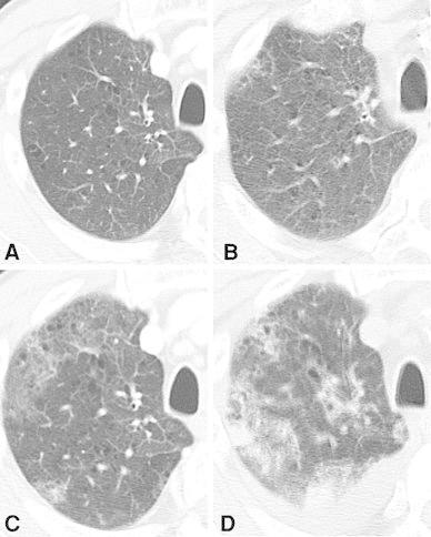
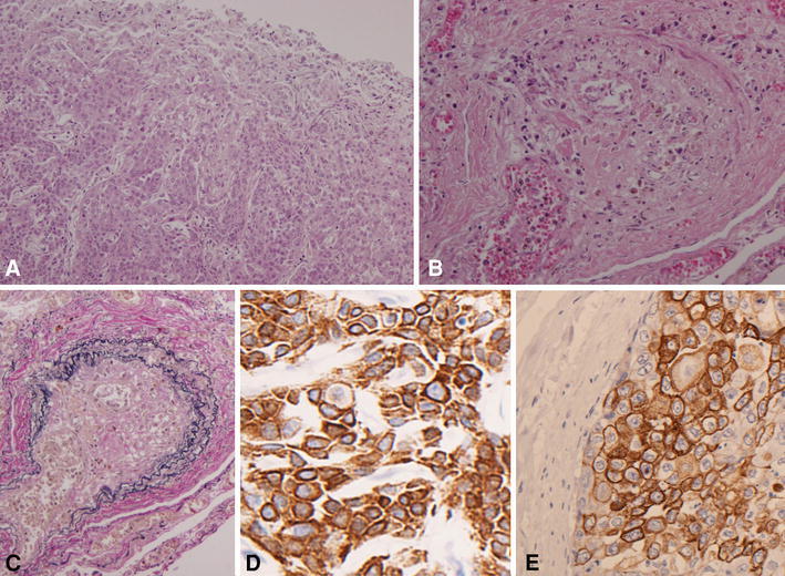
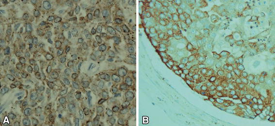
A 67-year-old male was referred to our hospital after being diagnosed with esophageal squamous cell carcinoma of the middle thoracic esophagus. The clinical stage was T1b(sm)N4M1 cStage IVb, so he was admitted to our hospital for systemic chemotherapy. He had sustained fever and a dry cough. Chest computed tomography showed the presence of irregular shadows, and unidentified respiratory insufficiency had progressed. A transbronchial lung biopsy revealed a pulmonary artery tumor embolus of esophageal squamous cell carcinoma. He developed DIC and died of respiratory failure on the 19th hospital day. The postmortem autopsy detected pulmonary tumor thrombotic microangiopathy accompanied by esophageal squamous cell carcinoma.

Keywords: Esophageal squamous cell carcinoma; PTTM; Pulmonary tumor thrombotic microangiopathy.


Figures

Fig. 1
Fig. 1
Esophageal squamous cell carcinoma was detected in the middle thoracic esophagus (a, b). Contrast-enhanced CT showed the multiple para-aortic lymph node metastases and cardiac right ventricle metastasis (c d). FDG-PET showed multiple high accumulations of FDG (shows SUV counts) (e)
Fig. 2
Fig. 2
The progressive multiple lung shadows were detected with chest CT. Twelve days before admission (a), on the 2nd hospital day (b), 11th day (c) and 19th day (d)
Fig. 3
Fig. 3
Microscopic examination. The primary tumor of the esophagus (a H&E ×100). Multiple tumor emboli and clot formation were shown in the pulmonary arterioles (b H&E ×100). EVG staining of the pulmonary arterioles showed concentric fibrocellular intimal proliferation with narrowing of the lumen (c EVG ×100). Immunostaining for CK5/6 proved that both the positivity rate and staining intensity were high (d primary tumor of the esophagus, CK5/6 ×400, e tumor cells in the pulmonary arteriole, CK5/6 ×400)
Fig. 4
Fig. 4
Immunostaining for VEGF. VEGF-positive tumor cells showed diffuse cytoplasmic staining (a primary tumor of the esophagus, VEGF ×400; b tumor cells in the pulmonary arteriole, VEGF ×400)

References

    1. von Harvey A, Illes A, Waldherr R, Otto H. Pulmonary tumor thrombotic microangiopathy with pulmonary hypertension. Cancer. 1990;66:587–592. doi: 10.1002/1097-0142(19900801)66:3<587::AID-CNCR2820660330>3.0.CO;2-J. - DOI - PubMed
    1. Japanese classification of esophageal cancer. The Japanese Society for Esophageal Diseases. 10th edn. 2008
    1. Kagata Y, Nakanishi K, Ozeki Y, Terahata S, Matsubara O. An immunohistochemical study of tumor thrombotic microangiopathy: the role of TF, FGF and VEGF. J Jpn coll Angiol. 2003;43:679–684.
    1. Hibbert M, Braude S. Tumor microembolism presenting as “primary pulmonary hypertension”. Thorax. 1997;52:1016–1017. doi: 10.1136/thx.52.11.1016. - DOI - PMC - PubMed
    1. Miyano S, Izumi S, Takeda Y, Tokuhara M, Mochizuki M, Matsubara O, et al. Pulmonary tumor thrombotic microangiopathy. J Clin Oncol. 2007;10:597–599. doi: 10.1200/JCO.2006.09.0670. - DOI - PubMed

LinkOut - more resources