. 2013 Oct 18;23(39):10.1002/adfm.201300570.
doi: 10.1002/adfm.201300570.

Highly Elastic Micropatterned Hydrogel for Engineering Functional Cardiac Tissue

Affiliations

Highly Elastic Micropatterned Hydrogel for Engineering Functional Cardiac Tissue

Nasim Annabi et al. Adv Funct Mater. .

Abstract

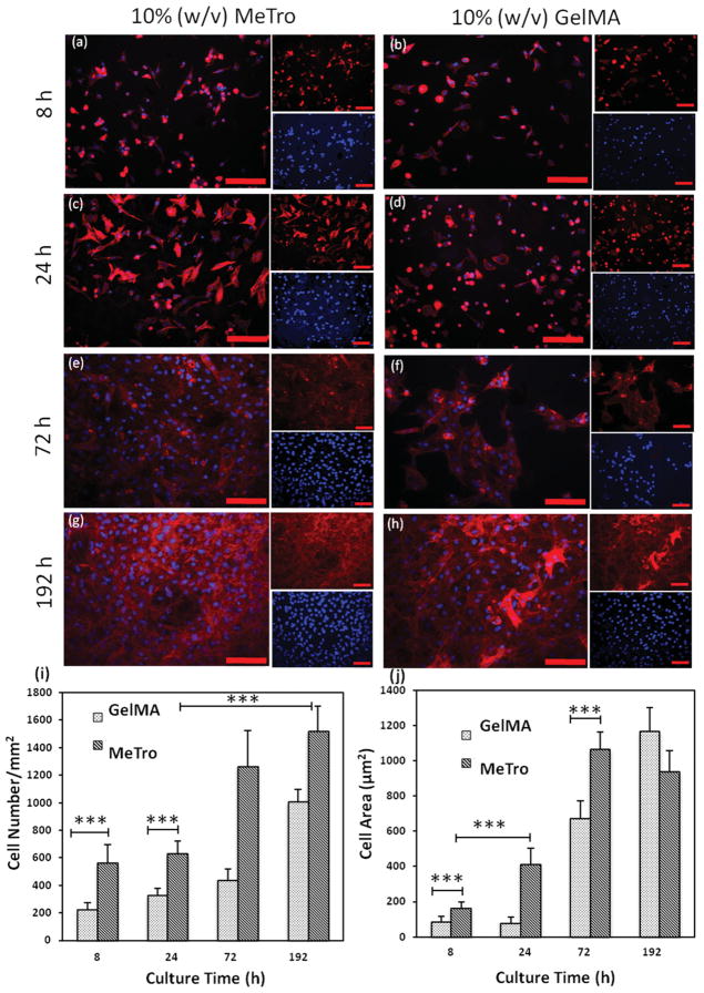
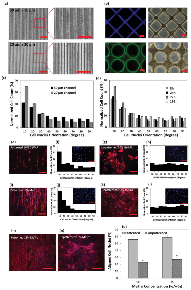
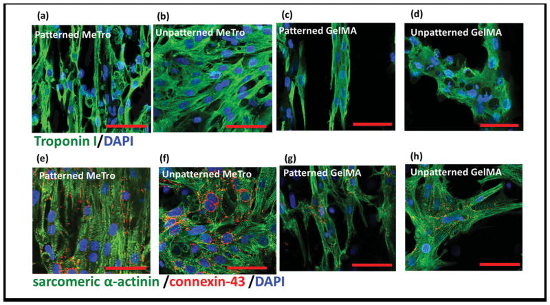
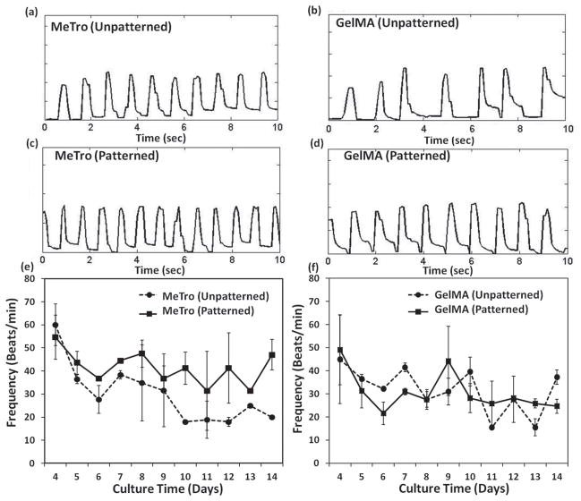
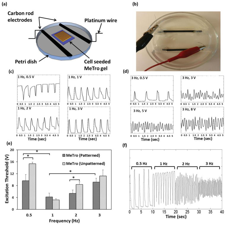
Heart failure is a major international health issue. Myocardial mass loss and lack of contractility are precursors to heart failure. Surgical demand for effective myocardial repair is tempered by a paucity of appropriate biological materials. These materials should conveniently replicate natural human tissue components, convey persistent elasticity, promote cell attachment, growth and conformability to direct cell orientation and functional performance. Here, microfabrication techniques are applied to recombinant human tropoelastin, the resilience-imparting protein found in all elastic human tissues, to generate photocrosslinked biological materials containing well-defined micropatterns. These highly elastic substrates are then used to engineer biomimetic cardiac tissue constructs. The micropatterned hydrogels, produced through photocrosslinking of methacrylated tropoelastin (MeTro), promote the attachment, spreading, alignment, function, and intercellular communication of cardiomyocytes by providing an elastic mechanical support that mimics their dynamic mechanical properties in vivo. The fabricated MeTro hydrogels also support the synchronous beating of cardiomyocytes in response to electrical field stimulation. These novel engineered micropatterned elastic gels are designed to be amenable to 3D modular assembly and establish a versatile, adaptable foundation for the modeling and regeneration of functional cardiac tissue with potential for application to other elastic tissues.


Conflict of interest statement

The authors declare no conflict of interest in this work.

Figures

Figure 1
Figure 1
Cardiomyocyte attachment and proliferation on 10% (w/v) MeTro and GelMA hydrogels as a function of time. Rhodamine-labeled phalloidin/ DAPI staining for F-actin/cell nuclei of cardiomyocytes seeded on MeTro and GelMA gels after a,b) 8 h, c,d) 24 h, e,f) 72 h, and g,h) 192 h of culture, demonstrating higher cell attachment and spreading on MeTro gels compared to GelMA at different culture times (smaller panels show F-actin (top) and cell nuclei (bottom) stained samples) (scale bar = 100 μm). i) Cell densities, defined as the number of DAPI stained nuclei per given hydrogel area, on MeTro and GelMA gels at various culture times. j) Cell spreading, defined as the area of cell clusters divided by the number of the cells within those cluster, on MeTro and GelMA hydrogels; the reduction in cell area on MeTro compared to GelMA gels after 192 h is a consequence of the higher cell proliferation on MeTro gels. Error bars represent the SD of measurements performed on 5 samples (***p < 0.001)
Figure 2
Figure 2
Cardiomyocyte elongation and alignment on the surface of micropatterned gels. Representative images of micropatterned MeTro, a) 20 × 20 μm and 50 × 50 μm channels (scale bar = 200 μm) produced by micromolding, b) gels of various geometry generated by photomasking (scale bar = 50 μm). c) Histogram of the relative alignment in 10° increments on day 3 of culture, demonstrating increased cellular alignment with decreasing microchannel width. d) Histogram of the relative alignment obtained at different culture time shows the highest cell alignment after 72 h of culture. Cell alignment on MeTro and GelMA hydrogels 72 h post seeding, representative F-actin/DAPI stained images with corresponding histograms of e,f) patterned GelMA and g,h) unpatterned GelMA; i,j) patterned MeTro; k,l) unpatterned MeTro, demonstrating higher cell alignment in 20° increments on micropatterned MeTro gel compared to patterned GelMA hydrogel (channel size: 20 × 20 μm, scale bar = 100 μm). Effect of MeTro concentration on cardiomyocyte elongation and alignment, representative F-actin/DAPI stained images of cardiomyocytes seeded on m) patterned and n) unpatterned MeTro gels produced by using 15% (w/v) MeTro, o) mean percentage of aligned cell nuclei (within 20° of preferred nuclear orientation), demonstrating cell alignment for both MeTro concentrations.
Figure 3
Figure 3
Immunostaining of cardiomyocyte markers. Hydrogels stained for a–d) troponin (green)/nuclei (blue) and e–h) sarcomeric α-actinin (green)/ connexin-43 (red)/nuclei (blue) on day 8 of culture, patterned MeTro gels are shown in (a) and (e) and unpatterned MeTro samples in (b) and (f), patterned GelMA in (c) and (g), and unpatterned GelMA in (d) and (h) (scale bar = 50 μm)
Figure 4
Figure 4
Beating characterization of cardiomyocytes seeded on hydrogels. Beating behavior of cardiomyocyte-seeded a,c) MeTro and b,d) GelMA gels on day 7 of culture, unpatterned samples are shown in (a,b) and patterned ones in (c,d). Spontaneous beating frequency of cardiomyocytes seeded on e) MeTro and f) GelMA gels over 14 days of culture.
Figure 5
Figure 5
Electrical stimulation of cell-seeded MeTro gels. a,b) Stimulation chamber for applying electrical stimuli to cardiomyocytes cultured on the surfaces of MeTro gels. Analyses of contractile response to electrical stimulation on day 8 of culture, voltage was gradually increased at c) 1 Hz and d) 3 Hz frequencies to induce synchronized beating, the beating became more synchronized by increasing the voltage. e) Excitation threshold of cardiac tissues on both patterned and unpatterned MeTro gels at various frequencies, demonstrating that the excitation threshold was lower in patterned MeTro gels compared to unpatterned ones. f) Recording of synchronous beating signals of cardiomyocytes cultured on patterned MeTro gels in response to applied external electric field at 0.5, 1, 2, and 3 Hz. Error bars represent the SD of measurements performed on 5 samples (*p < 0.05).
Scheme 1
Scheme 1
Schematic diagram describing the fabrication of micropatterned gels using a micro-molding process for cardiomyocyte alignment. a) Microchannels with varying channel sizes and spacings were formed on a silicon wafer, to produce a master, which was then covered with a layer of PDMS prepolymer. After curing for 1 h at 80 ° C, the PDMS mold was removed and cut into small molds (1 cm × 1 cm); b) a mold was then placed on a glass slide containing 10 μl MeTro and photoinitiator solution. Exposure to UV light for 35 s crosslinked the hydrogel and generated a micropatterned MeTro gel, the PDMS mold was removed from the gel after soaking in PBS for 5 min; c) the hydrogel containing microchannels was seeded with cardiomyocytes isolated from neonatal rats to align cells within the channels, an unpatterned MeTro gel was used as control.

References

    1. Rane AA, Christman KL. J Am Coll Cardiol. 2011;58:2615. - PubMed
    1. Bouten CVC, Dankers PYW, Driessen-Mol A, Pedron S, Brizard AMA, Baaijens FPT. Adv Drug Delivery Rev. 2011;63:221. - PubMed
    1. Radisic M, Park H, Gerecht S, Cannizzaro C, Langer R, Vunjak-Novakovic G. Philos Trans R Soc Lond B Biol Sci. 2007;362:1357. - PMC - PubMed
    1. Radisic M, Park H, Shing H, Consi T, Schoen FJ, Langer R, Freed LE, Vunjak-Novakovic G. Proc Natl Acad Sci USA. 2004;101:18129. - PMC - PubMed
    1. Miyagawa S, Roth M, Saito A, Sawa Y, Kostin S. Ann Thorac Surg. 2011;91:320. - PubMed