

Increased motor cortex excitability during motor imagery in brain-computer interface trained subjects
- PMID: 24319425
- PMCID: PMC3837244
- DOI: 10.3389/fncom.2013.00168
Increased motor cortex excitability during motor imagery in brain-computer interface trained subjects
Abstract
Background: Motor imagery (MI) is the mental performance of movement without muscle activity. It is generally accepted that MI and motor performance have similar physiological mechanisms.
Purpose: To investigate the activity and excitability of cortical motor areas during MI in subjects who were previously trained with an MI-based brain-computer interface (BCI).
Subjects and methods: Eleven healthy volunteers without neurological impairments (mean age, 36 years; range: 24-68 years) were either trained with an MI-based BCI (BCI-trained, n = 5) or received no BCI training (n = 6, controls). Subjects imagined grasping in a blocked paradigm task with alternating rest and task periods. For evaluating the activity and excitability of cortical motor areas we used functional MRI and navigated transcranial magnetic stimulation (nTMS).
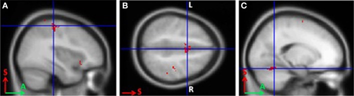
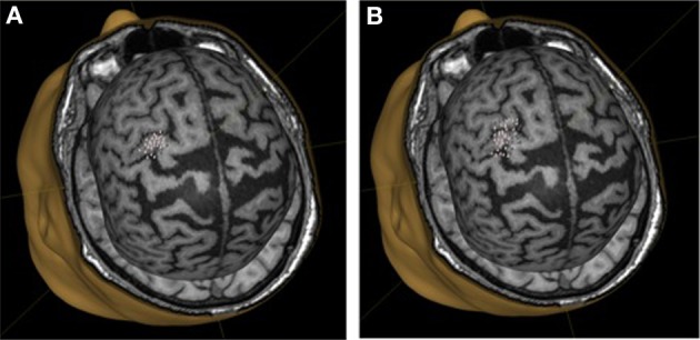
Results: fMRI revealed activation in Brodmann areas 3 and 6, the cerebellum, and the thalamus during MI in all subjects. The primary motor cortex was activated only in BCI-trained subjects. The associative zones of activation were larger in non-trained subjects. During MI, motor evoked potentials recorded from two of the three targeted muscles were significantly higher only in BCI-trained subjects. The motor threshold decreased (median = 17%) during MI, which was also observed only in BCI-trained subjects.
Conclusion: Previous BCI training increased motor cortex excitability during MI. These data may help to improve BCI applications, including rehabilitation of patients with cerebral palsy.
Keywords: brain-computer interface; functional MRI; motor imagery; navigated TMS; neurorehabilitation.
Figures


References
-
- Chervyakov A. V., Piradov M. A., Savitskaya N. G., Chernikova L. A., Kremneva E. I. (2013). New step to a personalized medicine. Navigation transcranial magnetic stimulation (nbs eximia nexstim). Ann. Clin. Exp. Neurol. 6, 37–46 [Article in Russian].
LinkOut - more resources
Full Text Sources
Other Literature Sources
Medical