

A case report of segmental arterial mediolysis in which computed tomography angiography was useful for diagnosis
- PMID: 24319500
- PMCID: PMC3851787
- DOI: 10.1007/s12328-013-0433-7
A case report of segmental arterial mediolysis in which computed tomography angiography was useful for diagnosis
Abstract
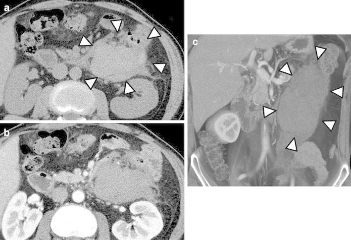
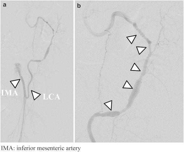
A 48-year-old male presented to our hospital with abdominal pain. Laboratory studies showed no abnormality, the severity of his abdominal pain decreased, and the patient was discharged. Five days later, the patient visited a neighborhood clinic because of fever with a 3-day history of temperatures of approximately 38 °C. The patient was admitted to our hospital 6 days after his initial visit. Laboratory investigation revealed a C-reactive protein level of 18.2 mg/dL. Abdominal computed tomography (CT) showed an 80 × 60 mm hematoma behind the descending colon, but no extravasation was detected. Thin-slice maximum-intensity-projection images from CT angiography (CTA) showed irregular narrowing and intermittent fusiform dilatations of the left colonic artery, suggesting a vascular disease, such as segmental arterial mediolysis (SAM). Digital subtraction angiography showed local irregularity, and 'beading and narrowing' of the left colonic artery, similar to the findings on CTA. Left hemicolectomy was electively performed on the twenty-fifth hospital day. Histological findings were consistent with SAM. Thus, CTA was a useful modality for the early diagnosis of SAM.
Keywords: CT angiography (CTA); Digital subtraction angiography (DSA); Maximum-intensity-projection (MIP) images; Segmental arterial mediolysis.
Figures


Similar articles
-
Segmental arterial mediolysis: CTA findings at presentation and follow-up.AJR Am J Roentgenol. 2006 Dec;187(6):1463-9. doi: 10.2214/AJR.05.0281. AJR Am J Roentgenol. 2006. PMID: 17114538
-
Left omental artery bleeding in two patients with segmental arterial mediolysis successfully isolated with coil embolization.CVIR Endovasc. 2020 Jul 19;3(1):36. doi: 10.1186/s42155-020-00127-0. CVIR Endovasc. 2020. PMID: 32686023 Free PMC article.
-
Clinically Suspected Segmental Arterial Mediolysis of the Splanchnic Arteries: A Report of 2 Rare Cases.Am J Case Rep. 2021 Apr 8;22:e929013. doi: 10.12659/AJCR.929013. Am J Case Rep. 2021. PMID: 33830972 Free PMC article.
-
Middle-colic artery aneurysm associated with segmental arterial mediolysis, successfully managed by transcatheter arterial embolization: report of a case.Surg Today. 2009;39(2):144-7. doi: 10.1007/s00595-008-3811-x. Epub 2009 Feb 7. Surg Today. 2009. PMID: 19198994 Review.
-
Segmental arterial mediolysis: a case of mistaken hemorrhagic pancreatitis and review of the literature.JOP. 2014 Jan 10;15(1):72-7. doi: 10.6092/1590-8577/2036. JOP. 2014. PMID: 24413790 Review.
Cited by
-
Longitudinal Evaluation of Segmental Arterial Mediolysis in Splanchnic Arteries: Case Series and Systematic Review.PLoS One. 2016 Aug 11;11(8):e0161182. doi: 10.1371/journal.pone.0161182. eCollection 2016. PLoS One. 2016. PMID: 27513466 Free PMC article.
-
Transarterial embolization for ruptured pancreaticoduodenal artery aneurysm due to segmental arterial mediolysis combined with median arcuate ligament syndrome: a case report.Clin J Gastroenterol. 2023 Dec;16(6):859-863. doi: 10.1007/s12328-023-01847-1. Epub 2023 Aug 22. Clin J Gastroenterol. 2023. PMID: 37608145
-
Spontaneous rupture of an intrahepatic aneurysm of the right hepatic artery caused by segmental arterial mediolysis.BMJ Case Rep. 2016 Mar 18;2016:bcr2015214109. doi: 10.1136/bcr-2015-214109. BMJ Case Rep. 2016. PMID: 26994049 Free PMC article.
-
Pancreaticoduodenal Artery Aneurysm Rupture Presenting as Duodenal Obstruction Successfully Treated with Early Transcatheter Arterial Embolization: A Case Report of Suspected Segmental Arterial Mediolysis.Intern Med. 2023 Dec 1;62(23):3479-3482. doi: 10.2169/internalmedicine.1278-22. Epub 2023 Apr 14. Intern Med. 2023. PMID: 37062731 Free PMC article.
References
-
- Slavin RE, Gonzalez-Vitale JC. Segmental mediolytic arteritis: a clinical pathologic study. Lab Invest. 1976;35(1):23–29. - PubMed
-
- Slavin RE, Saeki K, Bhagavan B, et al. Segmental arterial mediolysis: a precursor to fibromuscular dysplasia? Mod Pathol. 1995;8(3):287–294. - PubMed
-
- Ishizaki Y, Fukuda T, Nakahara M, et al. Two cases of intra-abdominal hematoma due to rupture of an abdominal visceral artery in which conservative therapy was possible. J Jpn Surg Assoc. 2008;69(4):776–780. doi: 10.3919/jjsa.69.776. - DOI
LinkOut - more resources
Full Text Sources
Other Literature Sources
Research Materials
