Review
. 2013:2013:853654.
doi: 10.1155/2013/853654. Epub 2013 Nov 11.

Translational approach to behavioral learning: lessons from cerebellar plasticity

Affiliations
Review

Translational approach to behavioral learning: lessons from cerebellar plasticity

Guy Cheron et al. Neural Plast. 2013.

Abstract

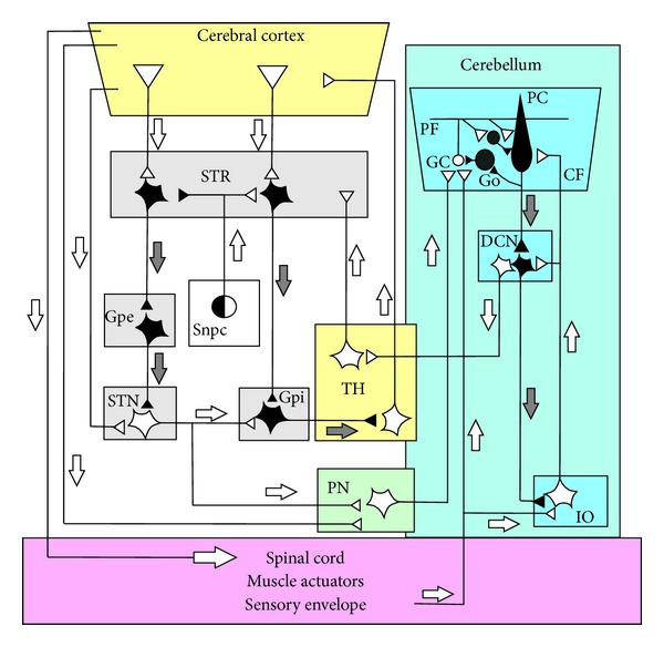
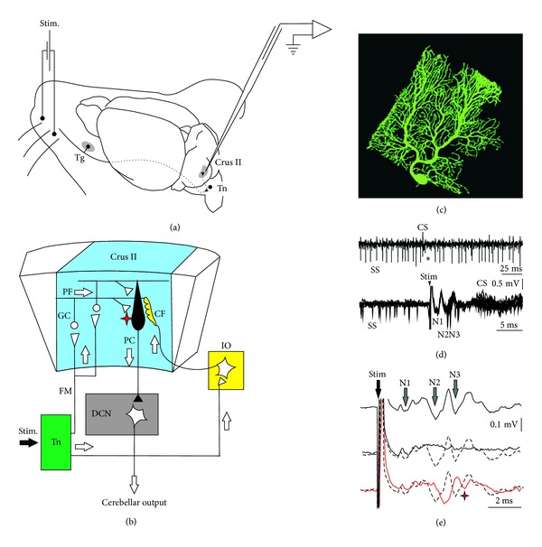
The role of cerebellar plasticity has been increasingly recognized in learning. The privileged relationship between the cerebellum and the inferior olive offers an ideal circuit for attempting to integrate the numerous evidences of neuronal plasticity into a translational perspective. The high learning capacity of the Purkinje cells specifically controlled by the climbing fiber represents a major element within the feed-forward and feedback loops of the cerebellar cortex. Reciprocally connected with the basal ganglia and multimodal cerebral domains, this cerebellar network may realize fundamental functions in a wide range of behaviors. This review will outline the current understanding of three main experimental paradigms largely used for revealing cerebellar functions in behavioral learning: (1) the vestibuloocular reflex and smooth pursuit control, (2) the eyeblink conditioning, and (3) the sensory envelope plasticity. For each of these experimental conditions, we have critically revisited the chain of causalities linking together neural circuits, neural signals, and plasticity mechanisms, giving preference to behaving or alert animal physiology. Namely, recent experimental approaches mixing neural units and local field potentials recordings have demonstrated a spike timing dependent plasticity by which the cerebellum remains at a strategic crossroad for deciphering fundamental and translational mechanisms from cellular to network levels.


Figures

Figure 1
Figure 1
Schematic diagram of the circuits interconnecting the olivocerebellum, the thalamus, the basal ganglia, the pontine nuclei, the cerebral cortex, and the spinal cord. The part of the circuit showing anatomical links between the basal-ganglia and the cerebellum is adapted from recent anatomical experiments using retrograde transneuronal transport of rabies virus from injections into the cerebellar cortex and in nuclei of basal ganglia, establishing evidence for disynaptic pathways that directly link the cerebellum with the basal ganglia (see [1] for a review). Injection of the rabies virus into the cerebellar cortex induced two stages of transport: retrograde transport to first-order neurons in the pontine nuclei (PN) that innervate the injection site and then retrograde transneuronal transport to second-order neurons in the subthalamic nucleus (STN) that innervate the first-order neurons [28]. Injection of rabies virus into the striatum (STR) induced retrograde transport to first-order neurons in the thalamus (TH) that innervate the injection site and then retrograde transneuronal transport to second-order neurons in the dentate nucleus (DN) that innervate the first-order neurons [29]. In addition, the striatal neurons that receive cerebellar inputs include neurons in the “indirect” pathway that send projections to the external globus pallidus (Gpe). The classical network of the basal ganglia (adapted from [30]) represented by the parallel “direct” and “indirect” pathways from the STR to the basal output nuclei. The “direct” path sent inhibitory input from the SRT to the internal part of the globus pallidus (Gpi). The basal ganglia circuit is completed by the dopaminergic pathway and represented the projection of the substantia nigra pars compacta (Snpc) to the STR. Inhibitory neurons are shown as filled symbols, excitatory neurons by open symbols. Grey and white arrows represent inhibitory or excitatory pathways, respectively. Abbreviations: PC, Purkinje cell; GO, Golgi cell; DCN, deep cerebellar nuclei; PF, parallel fiber; GC granule cell; IO, inferior olive; CF, climbing fiber.
Figure 2
Figure 2
VOR adaptation and Purkinje cell behavior.  (a) Schematic diagram of the circuits connecting the VOR pathway including the medial vestibular nucleus (MVN) and the neural integrator located in the prepositus hypoglossa nucleus (PH) and the horizontal microzone of the flocculus (Floc). The error signal initiated by the retinal slip is conveyed via the inferior olive (IO) by the climbing fiber (CF). The asterisk points to possible sites of VOR plasticity (see text for details). (b) Peristimulus time histograms of the complex spike (CS) and the simple spike (SS) responses of a representative Purkinje cell in the horizontal zone of the rabbit flocculus to sinusoidal rotation at 0.05 Hz in the light. Note the reciprocity of the CS and SS firing (adapted from   Figure  2 of [80] with permission). (c) Behavior of head velocity plus eye position sensitivity (HVplusP-P cell) recorded in the horizontal zone of the cat (adapted from Figure  3 of [24] with permission). (d) Time course of VOR adaptation corresponding to an out-of-phase VOR-OKN stimulation in 5 cats before (closed circles) and after (open circles) brainstem commissural incision. (e) Example of the VOR adaptation procedure used for increasing the VOR gain. Before the adaptation, the VOR is measured in the dark during table rotation (h) of 40°peak-to-peak at 0.10 Hz; the position of the eye (e) is recorded by means of the search coil technique. The VOR gain is the ratio between the peak-to-peak of the slow phase of the eye velocity and the peak-to-peak of the head velocity. During the training a random pattern of light circles was projected on the drum (d) surrounding the cat and oscillated out of phase of the head rotation, inducing an increase in the amplitude of the eye movements. After 4 hours of such training, the VOR was recorded in the dark and the VOR normalized gain increased by a mean gain of 1.41 ± 0.08 (adapted from Figures  1  and  2 of [77] with permission). Abbreviations: CsC, caudal semicircular canal; Floc, flocculus; Mn, motoneuron; OKN, optokinetic; LR, lateral rectus; VOR, vestibuloocular reflex.
Figure 3
Figure 3
Gilbert and Thach (1977) experiment in monkey.  (a) Task consist to control a handle horizontally by flexing or extending the wrist to a central position and to hold it there despite flexor and extensor loads applied to the handle. (b) CS and SS frequencies change for a PC after a change in load (horizontal arrow). Each grey bar represents an SS and the red dots a CS. Each row of bars represents the discharge during a trial successively represented from top to bottom. Each flexor trace on the left, for which the monkey performance was good, is followed by an extensor trace on the right. At the arrow, the known extensor load of 300 g was modified to a novel 450 g inducing a strong decrease in the performance. (c) Idealized representation of the SS and CS firing a long time and the number of trials before load perturbation (vertical arrow), during the transition period from bad to good performance, and after. (Adapted from [36, 98] with permission).
Figure 4
Figure 4
Directional learning during pursuit eye movements. (a) Representative eye movement during pursuit of a target moving first toward the right and then in oblique upward direction, the inset indicates the target motion in polar coordinates. (b) Eye velocity trace of the movement illustrated in (a). Gray and black traces indicate data from representative trials before learning and after at least 100 learning trials, respectively. The dashed traces indicate the velocity step signal of the target motion. Note that the vertical step (lower part) delayed the horizontal one (upper part) by 250 ms. The arrow pointing down and right indicates the learned response. The arrow pointing up and left indicates the hardwired visual response to the change in target direction, (adapted from [83] with permission).
Figure 5
Figure 5
Eyeblink  conditioning  protocols  and  electrophysiological  recording  of  the  conditioned  eyelid  response. (a) Experimental design for eyeblink conditioning in behaving rabbits illustrating the location of the bipolar hook electrodes for the recording of orbicularis oculi muscle activity (O.O. EMG). Classical eyeblink conditioning protocol consists of pairing a conditioned stimulus (CSt) (e.g., a neutral stimulus such as a tone) and an unconditioned stimulus (USt) (e.g., an airpuff to the eye that induces a reflexive blink). (b) Two principal paradigms have been classically used depending on the temporal relationship between CSt and USt. Thus, in the delay paradigm (top) the CSt and USt coterminate. In the trace paradigm there is a constant time interval between both stimuli (bottom). (c) The figure illustrates the eyeblink conditioning process using a delay paradigm. The conditioning paradigm (CS and US presentations) and representative orbicularis oculi electromyohraphic (O.O. EMG) recordings from the same animals along seven conditioning sessions (C1–C7) are presented. Along conditioning sessions the initial unconditioned response (UR), consisting of a reflexive eyelid response just after the US, leads to a timed eyelid response which precedes the USt named the conditioned response, CR (arrows).
Figure 6
Figure 6
Experimental design and electrophysiological response to electrical stimulation of mouse whiskers. (a) Animals were prepared for chronic recordings of local field potentials (LFP) and unitary extracellular activity in the Purkinje cell layer of the Crus I/II area. Facial dermatomes of the whisker region were electrically or tactilely stimulated with a pair of needles under the skin (Stim.) pulse, respectively. Sensory information comes into the Crus I/II area from the trigeminal nucleus (Tn) in the brainstem, which receives afferent signals from the trigeminal ganglion (Tg). (b) Schematic diagram of the circuits linking the trigeminal input and the olivo cerebellar system. (c) Photography of a single PC injected by Lucifer yellow. (d) Recording of spontaneous firing behavior of a Purkinje cell (PC) shows the presence of single spikes (SS) and complex spikes (CS). The presence of a CS followed by a pause in the SS firing (asterisk) identifies this neuron as a PC. Single trials, superimposed (n = 11), show spontaneous firing before the whisker electrical stimulation (Stim) and the temporal reorganization of the firing after the stimulus. SS firing occurred at the low points of the N2 and N3 components and later. The evoked CS occurred at a latency of 9–13 ms after the stimulus onset (arrowhead). (e) Long-term depression on the LFP is evident after the 8 Hz stimulation protocol (red line), when the latency of the N2 components increased and the amplitude of the N3 component strongly decreased. These effects were maximal just after the 8 Hz stimulation and persisted for at least 30 min. Single traces selected to compare latencies and amplitudes of LFP components; (top) before 8 Hz stimulation protocol, (middle) during 8 Hz stimulation, and (bottom) just after 8 Hz stimulation protocol. The red asterisk indicates the LTD effect on N3 postsynaptic components after 8 Hz stimulation, (adapted from [37] with permission).

References

    1. Bostan AC, Dum RP, Strick PL. Cerebellar networks with the cerebral cortex and basal ganglia. Trends in Cognitive Sciences. 2013;17(5):241–254. - PMC - PubMed
    1. Delgado-García JM, Gruart A. The role of interpositus nucleus in eyelid conditioned responses. Cerebellum. 2002;1(4):289–308. - PubMed
    1. Delgado-García JM, Gruart A. Building new motor responses: eyelid conditioning revisited. Trends in Neurosciences. 2006;29(6):330–338. - PubMed
    1. Medina JF, Nores WL, Mauk MD. Inhibition of climbing fibres is a signal for the extinction of conditioned eyelid responses. Nature. 2002;416(6878):330–333. - PubMed
    1. Hikosaka O, Nakamura K, Sakai K, Nakahara H. Central mechanisms of motor skill learning. Current Opinion in Neurobiology. 2002;12(2):217–222. - PubMed

Publication types

LinkOut - more resources