. 2013 Sep 1;2(9):e26246.
doi: 10.4161/onci.26246. Epub 2013 Sep 26.

A CD38/CD203a/CD73 ectoenzymatic pathway independent of CD39 drives a novel adenosinergic loop in human T lymphocytes

Affiliations

A CD38/CD203a/CD73 ectoenzymatic pathway independent of CD39 drives a novel adenosinergic loop in human T lymphocytes

Alberto L Horenstein et al. Oncoimmunology. .

Abstract

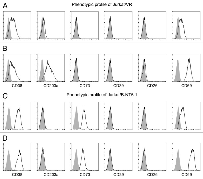
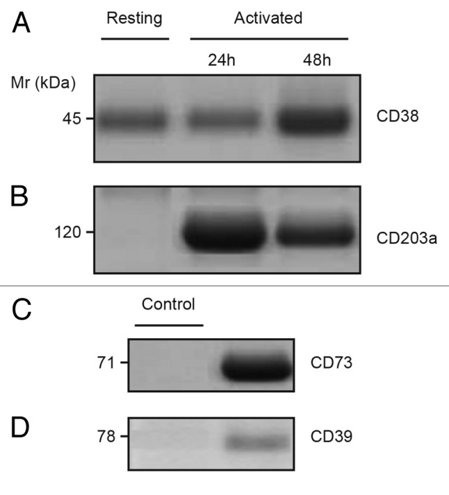
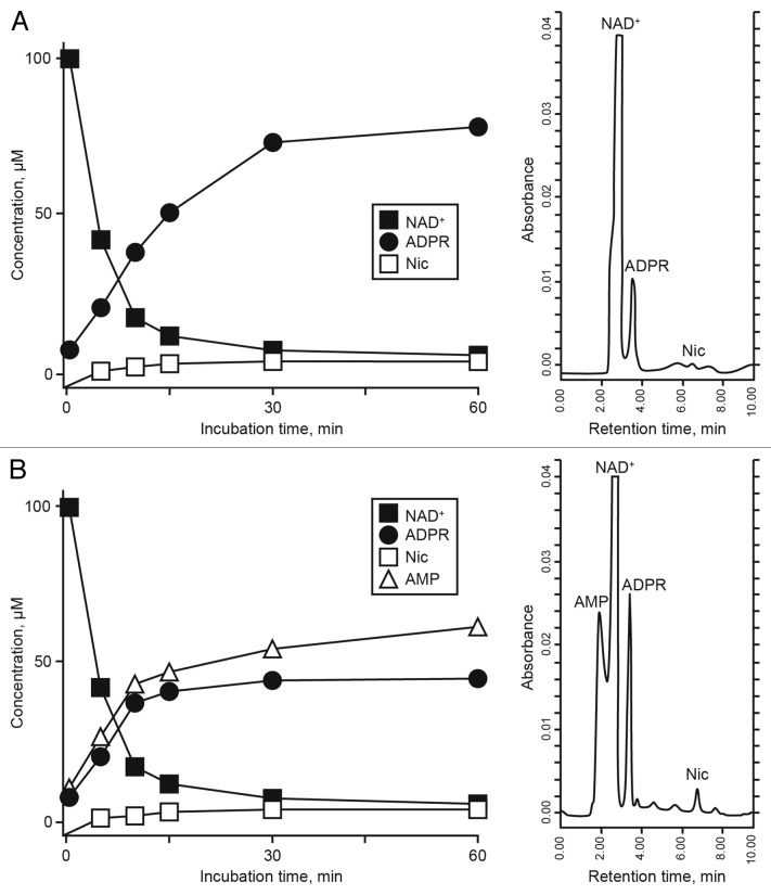
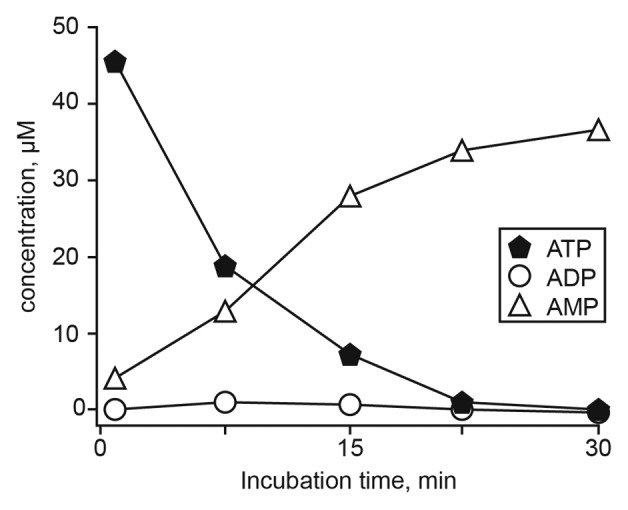
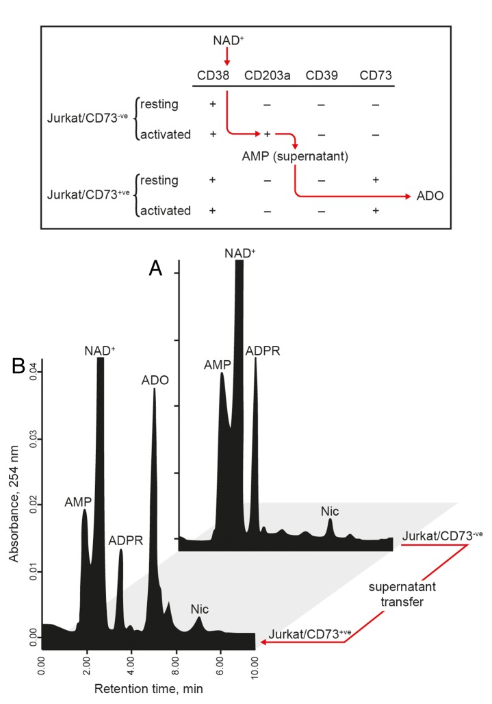
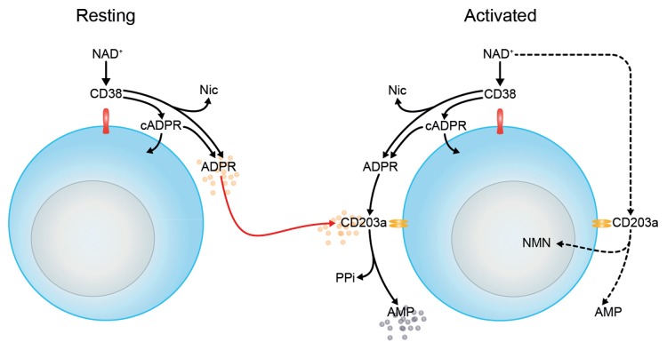
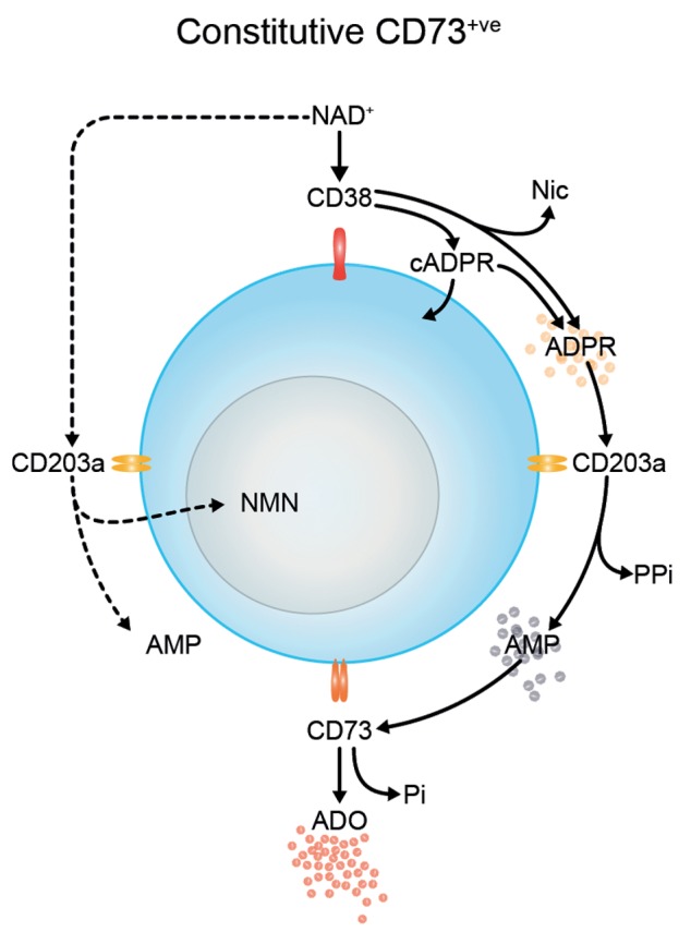
The tumor microenvironment is characterized by of high levels of extracellular nucleotides that are metabolized through the dynamic and sequential action of cell surface enzymes (ectoenzymes). These ectoenzymes operate according to their spatial arrangement, as part of (1) continuous (molecules on the same cell) or (2) discontinuous (molecules on different cells) pathways, the latter being facilitated by restricted cellular microenvironment. The outcome of this catabolic activity is an increase in the local concentration of adenosine, a nucleoside involved in the control of inflammation and immune responses. The aim of the work presented here was to demonstrate that a previously unexplored enzymatic pathway may be an alternate route to produce extracellular adenosine. Our data show that this new axis is driven by the nucleotide-metabolizing ectoenzymes CD38 (an NAD+ nucleosidase), the ecto-nucleotide pyrophosphatase/phosphodiesterase 1 (NPP1, also known as CD203a or PC-1) and the 5' ectonucleotidase (5'-NT) CD73, while bypassing the canonical catabolic pathway mediated by the nucleoside tri- and diphosphohydrolase (NTPDase) CD39. To determine the relative contributions of these cell surface enzymes to the production of adenosine, we exploited a human T-cell model allowing for the modular expression of the individual components of this alternative pathway upon activation and transfection. The biochemical analysis of the products of these ectoenzymes by high-performance liquid chromatography (HPLC) fully substantiated our working hypothesis. This newly characterized pathway may facilitate the emergence of an adaptive immune response in selected cellular contexts. Considering the role for extracellular adenosine in the regulation of inflammation and immunogenicity, this pathway could constitute a novel strategy of tumor evasion, implying that these enzymes may represent ideal targets for antibody-mediated therapy.

Keywords: CD203a; CD38; NAD+; PC-1; adenosine; ectonucleotidases.


Figures

None
Figure 1. Expression of ectoenzymes on activated and resting Jurkat cell lines. (A-D) Comparative cytofluorometric analysis of Jurkat T cells stained with primary antibodies against various ectonucleotidases and detected with fluorescein (FITC)-conjugated secondary antibodies. Expression analysis of ectoenzymes (CD38, CD203a, CD73, CD39, and CD26) and T-cell activation marker (CD69) by Jurkat/CD73 (A and B) and Jurkat/CD73+ (C and D) cells either in resting conditions (A and C) or in response to phorbol 12-myristate 13-acetate (B and D). Representative histograms are shown. Grey peaks demarcate isotype control staining and black peaks depict the expression levels of the indicated markers.
None
Figure 2. Constitutive expression of CD38 and elevated CD203a in activated Jurkat/CD73 cells. (A–B) Immunoblotting analysis of plasma membrane fractions from resting and phorbol 12-myristate 13-acetate (PMA)-activated Jurkat/CD73- cells probed with anti-CD38 (SUN-4B7, IgG1) mAbs or with anti-CD203a (3E8, IgG1) to detect the presence of 45-kDa CD38 (A) and 120-kDa CD203a (B) proteins. (C–D) Proteins with Mr of 71-kDa and 78-kDa, not expressed by Jurkat T cells (not shown), were immunodetected using lysates of epithelial cells from biopsied corneas used as positive controls and probed with anti-CD73 (CB73, IgG1) and anti-CD39 (IgG1) mAbs (right lanes). Isotype-matched irrelevant X63.Ag8 mAb was used as negative controls (left lanes). Mr = molecular weight”.
None
Figure 3. Analysis of NAD+ metabolites in the supernatant of Jurkat/CD73 cells. (A and B) The enzymatic conversion of NAD+ to adenosine diphosphate ribose (ADPR) (a CD38-dependent reaction) and to AMP (CD203a-dependent) was monitored by high-pressure liquid chromatography (HPLC). Representative resting (A) and phorbol 12-myristate 13-acetate (PMA)-activated-activated (B) Jurkat/CD73 cells upon incubation for 60 min at 37°C with 100 µM NAD+ are shown. The identity of peaks was confirmed by the co-migration and absorbance spectra of reference standards using a retention time (Rt) window of ± 5%. A representative plot of retention time of NAD+ metabolites in a single HPLC run is depicted on the right.
None
Figure 4. Time course of extracellular ATP metabolism in activated Jurkat/CD73 cells. ATP (100 μM) was added at time 0 and samples (500 µL) collected from the supernatants at the indicated times (abscissa). Sample were analyzed by HPLC to quantify ATP, ADP and AMP. The identity of peaks was confirmed by the co-migration and absorbance spectra of reference standards using a retention time (Rt) window of ± 5%. Vertical bars depicting the SEM do not exceed the size of symbols.
None
Figure 5. Metabolism of extracellular AMP consumption in Jurkat/CD73+ cells. (A-C) Jurkat/CD73+ cells were incubated with 100 µM AMP and the consequent (CD73-dependent) production of adenosine (ADO) was determined by HPLC. The identity of peaks was confirmed by the co-migration and absorbance spectra of reference standards using a retention time (Rt) window of ± 5%. (A) Experiments at t = 5 min and after 30 min incubation with 100 µM AMP. (B) AMP consumption in Jurkat/CD73+ cells exposed (solid symbols) or not exposed (open symbols) to 50 µM α,β-methylene-ADP (APCP). (C) Catabolism of AMP to ADO in the presence (solid symbols) or in the absence (open symbols) of 50 µM APCP. Representative Rt plots of metabolites in a single HPLC run are shown.
None
Figure 6. Jurkat T cells generates adenosine from NAD+. (A) The enzymatic conversion of NAD+ to AMP (a CD38/CD203a-dependent reaction) and to adenosine (ADO) (a CD73-dependent reaction) was evaluated. Activated Jurkat/CD73 cells were incubated for 60 min in buffer supplemented with 100 µM NAD+ and the resulting supernatant transferred to Jurkat/CD73+ cells, followed by further incubation for 30 min. (B) Supernatants from each step depicted in (A) were analyzed by HPLC for the presence of NAD+ catabolites. The identity of peaks was confirmed by the co-migration and absorbance spectra of reference standards using a retention time (Rt) window of ± 5%. Representative Rt plots of metabolites in a single HPLC run are shown.
None
Figure 7. CD38/CD203a ectoenzyme tandem catalytic cascade to degrade NAD+ in activated Jurkat/CD73 cells. Extracellular NAD+ is metabolized by the nicotinamide adenine dinucleotidase (NADase) CD38 expressed by resting Jurkat cells, generating nicotinamide (Nic), cyclic adenosine diphosphate ribose (cADPR) and ADPR. The latter product is transformed to AMP by the nucleotide pyrophosphatase/phosphodiesterase (NPP) CD203a expressed by phorbol 12-myristate 13-acetate (PMA)-activated Jurkat cells.
None
Figure 8. Biochemical activity of the CD38/CD203a/CD73 pathway in Jurkat T cells upon activation or transgene-driven CD73 expression. Extracellular NAD+ is metabolized by the nicotinamide adenine dinucleotidase (NADase) CD38, generating nicotinamide (Nic), cyclic adenosine diphosphate ribose (cADPR) and ADPR. The latter compound is transformed to AMP by nucleotide pyrophosphatase/phosphodiesterase (NPP) CD203a and then converted to adenosine (ADO) and inorganic pyrophosphate (PPi) by 5′-nucleotidase (5′NT) CD73. NMN, nicotinamide mononucleotide.

References

    1. Lanier LL. Shades of grey--the blurring view of innate and adaptive immunity. Nat Rev Immunol. 2013;13:73–4. doi: 10.1038/nri3389. - DOI - PubMed
    1. Kirschner MW. The meaning of systems biology. Cell. 2005;121:503–4. doi: 10.1016/j.cell.2005.05.005. - DOI - PubMed
    1. Malavasi F, Deaglio S, Funaro A, Ferrero E, Horenstein AL, Ortolan E, Vaisitti T, Aydin S. Evolution and function of the ADP ribosyl cyclase/CD38 gene family in physiology and pathology. Physiol Rev. 2008;88:841–86. doi: 10.1152/physrev.00035.2007. - DOI - PubMed
    1. Zimmermann H, Zebisch M, Sträter N. Cellular function and molecular structure of ecto-nucleotidases. Purinergic Signal. 2012;8:437–502. doi: 10.1007/s11302-012-9309-4. - DOI - PMC - PubMed
    1. Grahnert A, Grahnert A, Klein C, Schilling E, Wehrhahn J, Hauschildt S. Review: NAD+: a modulator of immune functions. Innate Immun. 2011;17:212–33. doi: 10.1177/1753425910361989. - DOI - PubMed

Publication types