. 2013 Aug 22;1(3):209-17.
doi: 10.1016/j.stemcr.2013.07.003. eCollection 2013.

Robust self-renewal of rat embryonic stem cells requires fine-tuning of glycogen synthase kinase-3 inhibition

Affiliations

Robust self-renewal of rat embryonic stem cells requires fine-tuning of glycogen synthase kinase-3 inhibition

Yaoyao Chen et al. Stem Cell Reports. .

Abstract

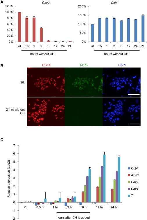
Germline-competent embryonic stem cells (ESCs) have been derived from mice and rats using culture conditions that include an inhibitor of glycogen synthase kinase 3 (GSK3). However, rat ESCs remain susceptible to sporadic differentiation. Here, we show that unsolicited differentiation is attributable to overinhibition of GSK3. The self-renewal effect of inhibiting GSK3 is mediated via β-catenin, which abrogates the repressive action of TCF3 on core pluripotency genes. In rat ESCs, however, GSK3 inhibition also leads to activation of differentiation-associated genes, notably lineage specification factors Cdx2 and T. Lowered GSK3 inhibition reduces differentiation and enhances clonogenicity and self-renewal. The differential sensitivity of rat ESCs to GSK3 inhibition is linked to elevated expression of the canonical Wnt pathway effector LEF1. These findings reveal that optimal GSK3 inhibition for ESC propagation is influenced by the balance of TCF/LEF factors and can vary between species.


Figures

None
Graphical abstract
Figure 1
Figure 1
Differentiation and Ectopic Expression of CDX2 in Rat ESCs (A) Bright-field and immunofluorescence images of rat ESCs in 2iL on feeders. (B) Immunostaining of OCT4 and CDX2 in mouse (mES) and rat (rES) ESCs. (C) Comparative analysis by qRT-PCR of Oct4, Nanog, and Cdx2 transcripts in mouse (blue) and rat (orange) ESCs using primers designed against conserved sequences. Expression values are normalized to Gapdh and relative to the average of mouse samples. Data were analyzed by unpaired t test. p < 0.01. (D) Immunostained rat E5.5 blastocyst. (E) qRT-PCR analysis of Fgfr2, Elf4, Eomes, Oct4, and Cdx2 in rESCs in 2iL (blue) and rat embryonic day 5.5 (E5.5) whole blastocysts (red line). Values are normalized to Gapdh and relative to the average in rat blastocysts. Error bars are SD of technical triplicates. Scale bar, 100 μM.
Figure 2
Figure 2
Effect of GSK3 Inhibition on Cdx2 Expression (A) Expression of Cdx2 and Oct4 upon CH removal. Values are normalized to Gapdh and relative to 2iL. (B) Immunofluorescence for CDX2 and OCT4 in rat ESCs cultured in 2iL and 24 hr after CH removal. (C) Transcriptional response of rat ESCs to CH. Expression is normalized to Gapdh and relative to values in PL. Error bars are SD of technical triplicates. Scale bar, 100 μM.
Figure 3
Figure 3
Titration of GSK3 Inhibition (A) qRT-PCR analysis of gene expression in rat ESCs cultured with different concentrations of CH. Values are normalized to Gapdh and relative to 2iL. Error bars represent SD of three technical replicates. (B and C) Immunofluorescence staining of rat ESCs cultured in T2iL or 2iL for CDX2 and T, respectively. (D) Morphology of rat ESC bulk cultures. (E) Colony formation from 250 single cells analyzed by AP staining. Error bars represent SD of four technical replicates. (F) Chimeras and germline F1 pups from injection of DA (Agouti) rat ESCs into SD (albino) blastocysts. Scale bar, 100 μM.
Figure 4
Figure 4
Interrogating Downstream Effectors of GSK3 Inhibition in Rat ESCs (A) TopFlash assay of β-catenin transcriptional activity in rat and mouse ESCs in PL, T2iL, and 2iL. Values are normalized to mouse ESCs in PL. Error bars represent SD of technical triplicates. (B) Immunostaining of β-catenin in rat ESCs cultured in T2iL and 2iL. (C) qRT-PCR analysis of Tcf1, Tcf3, Tcf4, and Lef1 in rat ESCs maintained in T2iL, using TaqMan probes. Values are normalized to Gapdh and relative to Tcf1. (D) qRT-PCR analysis using conserved primers of Lef1 expression in rat and mouse ESCs maintained in 2iL. (E) qRT-PCR analysis of a panel of gene expression after Tcf3 and Lef1 knockdown. Gene expression was normalized to Gapdh and relative to values in siGFP transfected cells cultured in 2iL. Error bars represent SD of technical triplicates. (F) Effect of stable knockdown of LEF1 on colony formation in 2iL or T2iL. A total of 80 cells were plated per well and analyzed by AP staining after 5 days. Error bars are SDs of four technical replicates. Data were analyzed by unpaired t test. p < 0.01; ∗∗p < 0.001. (G) Bright-field and immunofluorescence images of rat ESCs in 2iL with or without Lef1 knockdown. Scale bar, 100 μM.

References

    1. Beck F., Erler T., Russell A., James R. Expression of Cdx-2 in the mouse embryo and placenta: possible role in patterning of the extra-embryonic membranes. Dev. Dyn. 1995;204:219–227. - PubMed
    1. Berg D.K., Smith C.S., Pearton D.J., Wells D.N., Broadhurst R., Donnison M., Pfeffer P.L. Trophectoderm lineage determination in cattle. Dev. Cell. 2011;20:244–255. - PubMed
    1. Blair K., Wray J., Smith A. The liberation of embryonic stem cells. PLoS Genet. 2011;7:e1002019. - PMC - PubMed
    1. Blair K., Leitch H.G., Mansfield W., Dumeau C.É., Humphreys P., Smith A.G. Culture parameters for stable expansion, genetic modification and germline transmission of rat pluripotent stem cells. Biol. Open. 2012;1:58–65. - PMC - PubMed
    1. Bone H.K., Nelson A.S., Goldring C.E., Tosh D., Welham M.J. A novel chemically directed route for the generation of definitive endoderm from human embryonic stem cells based on inhibition of GSK-3. J. Cell Sci. 2011;124:1992–2000. - PMC - PubMed

Publication types

MeSH terms