. 2013 Dec 9;20(1):91.
doi: 10.1186/1423-0127-20-91.

Tonotopic reorganization and spontaneous firing in inferior colliculus during both short and long recovery periods after noise overexposure

Affiliations

Tonotopic reorganization and spontaneous firing in inferior colliculus during both short and long recovery periods after noise overexposure

Feng Wang et al. J Biomed Sci. .

Abstract

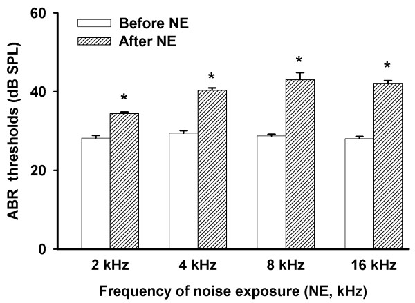
Background: Noise induced injury of the cochlea causes shifts in activation thresholds and changes of frequency response in the inferior colliculus (IC). Noise overexposure also induces pathological changes in the cochlea, and is highly correlated to hearing loss. However, the underlying mechanism has not been fully elucidated. In this study, we hypothesized that overexposure to noise induces substantial electrophysiological changes in the IC of guinea pigs.

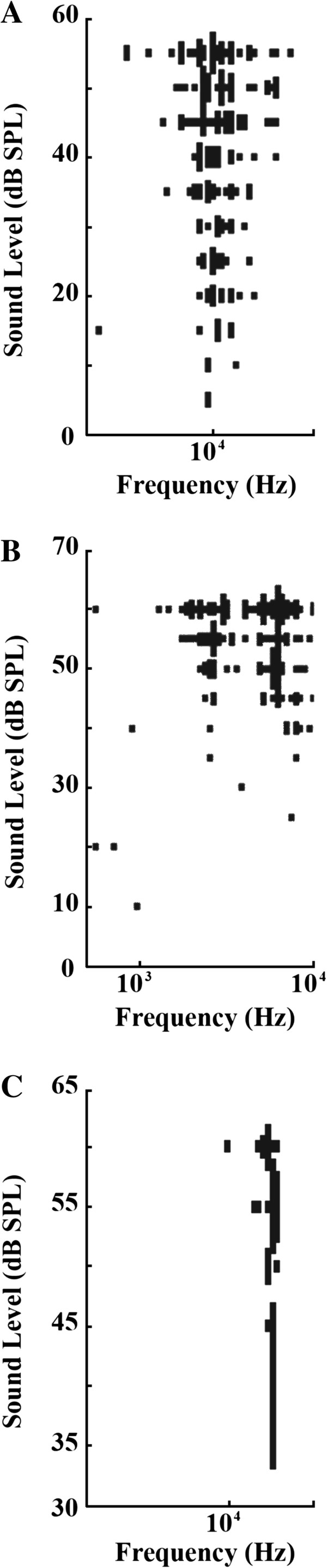
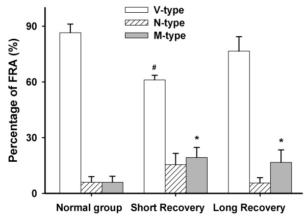
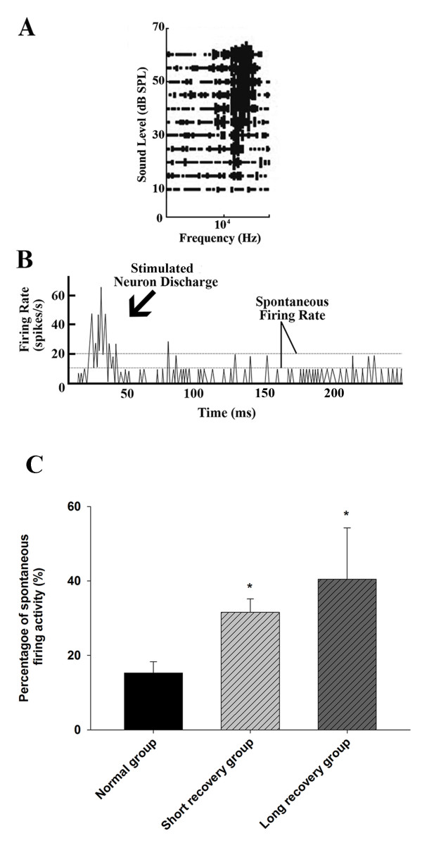
Results: During the noise exposure experiment, the animals were undergoing a bilateral exposure to noise. Additionally, various techniques were employed including confocal microscopy for the detection of cochlea hair cells and single neuron recording for spontaneous firing activity measurement. There were alterations among three types of frequency response area (FRA) from sound pressure levels, including V-, M-, and N-types. Our results indicate that overexposure to noise generates different patterns in the FRAs. Following a short recovery (one day after the noise treatment), the percentage of V-type FRAs considerably decreased, whereas the percentage of M-types increased. This was often caused by a notch in the frequency response that occurred at 4 kHz (noise frequency). Following a long recovery from noise exposure (11-21 days), the percentage of V-types resumed to a normal level, but the portion of M-types remained high. Interestingly, the spontaneous firing in the IC was enhanced in both short and long recovery groups.

Conclusion: Our data suggest that noise overexposure changes the pattern of the FRAs and stimulates spontaneous firing in the IC in a unique way, which may likely relate to the mechanism of tinnitus.


Figures

Figure 1
Figure 1
ABR Thresholds of auditory brainstem response before and after noise exposure (NE). Statistical data representative of ABR (auditory brainstem response) thresholds before and one day after noise exposure (n = 13 from 13 animals) in guinea pigs. Four frequencies were tested: 2 kHz, 4 kHz, 8 kHz, and 16 kHz. *Significant difference from pre-NE group at the same frequency (P < 0.01).
Figure 2
Figure 2
Representative confocal microscopy (x 40) of fluorescence stained nuclei within inner and outer hair cells in the cochlea of guinea pigs, 11 days after noise exposure (120 dB, 4 hours). A: Normal levels are observed in the nuclei of inner and outer hair cells of the control subject. The nuclei were arranged normally and no disruption or fragmentations were found. B: The image location is in the third turn of the cochlea after noise exposure, where signs of fragmentation equally occurred throughout both inner and outer hair cells. C: In the second turn of the cochlea, after noise exposure, more disruption and fragmentation appears to occur in the inner and outer hair cell nuclei compared to the third turn of the cochlea. D: The first turn in the cochlea, after noise exposure, exhibited severe disruption and fragmentation compared to the second and third turns in the inner and outer hair cell nuclei.
Figure 3
Figure 3
The number of nuclei in the cochlea in inner (A) and outer (B) hair cells. Before noise exposure (NE) vs. after NE (n = 12 from 6 animals; * P < 0.05).
Figure 4
Figure 4
Representative graphs of frequency response patterns that are the basis of FRA types. (A) V-type, (B) M-type, (C) and N-type FRAs created by testing neuronal responses through electrode measurement. Variables included sound frequency and intensity.
Figure 5
Figure 5
The percentage of FRA types including V-, N- and M-types in normal, short recovery, and long recovery groups. # P < 0.05 compared to normal groups in V-type (n = 6 from 6 animals for normal and n = 7 from 7 animals for short recovery). * P < 0.05 compared to normal groups in M-type (n = 6 from 6 animals for normal, n = 7 from 7 animals for short recovery, and n = 6 from 6 animals for long recovery).
Figure 6
Figure 6
Function curves providing a correlation between CF (characteristic frequency) and protrusion depth within the IC. A: A normal distribution can be observed within the control CF map. CF and depth have a positive trend, resulting in higher frequencies the deeper the protrusions within the IC. B: Short recovery group shows more of a randomized pattern with dispersed responses above and below 4 kHz creating a gap specifically at that frequency. C: Long recovery group exhibited the same type of gap at 4 kHz, but at a reduced level showing signs of recovery.
Figure 7
Figure 7
Spontaneous firing activities in IC neurons. A: Representative FRA showing typical spontaneous firing. B: A typical spontaneous firing curve over time in an IC neuron. C: The percentage of neurons with spontaneous firing activity in normal (114 neurons from 6 animals), short recovery (161 neurons from 7 animals) and long recovery groups (74 neuron from 6 animals). *Significant difference from normal group (P < 0.05).

Similar articles

Cited by

References

    1. Bauer CA, Turner JG, Caspary DM, Myers KS, Brozoski TJ. Tinnitus and inferior colliculus activity in chinchillas related to three distinct patterns of cochlear trauma. J Neurosci Res. 2008;20:2564–2578. doi: 10.1002/jnr.21699. - DOI - PMC - PubMed
    1. Dong S, Mulders WH, Rodger J, Woo S, Robertson D. Acoustic trauma evokes hyperactivity and changes in gene expression in guinea-pig auditory brainstem. Eur J Neurosci. 2010;20:1616–1628. - PubMed
    1. Mulders WH, Robertson D. Hyperactivity in the auditory midbrain after acoustic trauma: dependence on cochlear activity. Neuroscience. 2009;20:733–746. doi: 10.1016/j.neuroscience.2009.08.036. - DOI - PubMed
    1. Alkhatib A, Biebel UW, Smolders JW. Reduction of inhibition in the inferior colliculus after inner hair cell loss. Neuroreport. 2006;20:1493–1497. doi: 10.1097/01.wnr.0000234754.11431.ee. - DOI - PubMed
    1. Rajan R. Receptor organ damage causes loss of cortical surround inhibition without topographic map plasticity. Nat Neurosci. 1998;20:138–143. doi: 10.1038/388. - DOI - PubMed

Publication types