Review
. 2013 Dec 9;20(1):92.
doi: 10.1186/1423-0127-20-92.

DNA transposon-based gene vehicles - scenes from an evolutionary drive

Affiliations
Review

DNA transposon-based gene vehicles - scenes from an evolutionary drive

Kristian Alsbjerg Skipper et al. J Biomed Sci. .

Abstract

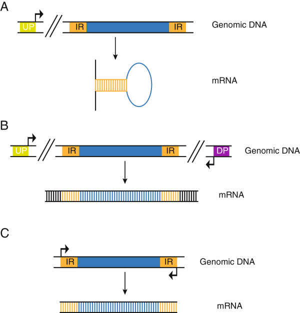
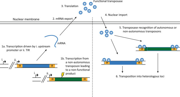
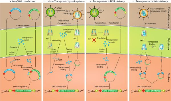
DNA transposons are primitive genetic elements which have colonized living organisms from plants to bacteria and mammals. Through evolution such parasitic elements have shaped their host genomes by replicating and relocating between chromosomal loci in processes catalyzed by the transposase proteins encoded by the elements themselves. DNA transposable elements are constantly adapting to life in the genome, and self-suppressive regulation as well as defensive host mechanisms may assist in buffering 'cut-and-paste' DNA mobilization until accumulating mutations will eventually restrict events of transposition. With the reconstructed Sleeping Beauty DNA transposon as a powerful engine, a growing list of transposable elements with activity in human cells have moved into biomedical experimentation and preclinical therapy as versatile vehicles for delivery and genomic insertion of transgenes. In this review, we aim to link the mechanisms that drive transposon evolution with the realities and potential challenges we are facing when adapting DNA transposons for gene transfer. We argue that DNA transposon-derived vectors may carry inherent, and potentially limiting, traits of their mother elements. By understanding in detail the evolutionary journey of transposons, from host colonization to element multiplication and inactivation, we may better exploit the potential of distinct transposable elements. Hence, parallel efforts to investigate and develop distinct, but potent, transposon-based vector systems will benefit the broad applications of gene transfer. Insight and clever optimization have shaped new DNA transposon vectors, which recently debuted in the first DNA transposon-based clinical trial. Learning from an evolutionary drive may help us create gene vehicles that are safer, more efficient, and less prone for suppression and inactivation.


Figures

Figure 1
Figure 1
Model of cut-and-paste transposition. Transposase proteins (green spheres) recognize the terminal inverted repeats (IRs, orange boxes) and form a circular pre-excision synaptic complex from which the transposon is excised. Formation of the synaptic complex, allowing close association of the two transposon ends, involves the formation of transposase tetramers [74]. Subsequently, the transposition complex recognises a target site and the transposase proteins mediate integration of the transposon. The figure shows the proposed mechanisms for Tc1/mariner-type (e.g. Sleeping Beauty) DNA transposition. Yellow boxes marked with IR represent the terminal inverted repeats.
Figure 2
Figure 2
Schematic representation of cut-and-paste transposition. (A) Transposition of Tc1/mariner elements (like Sleeping Beauty and Frog Prince) leads to double-stranded breaks and formation of a 2 or 3 bp 3′-overhang at the excision site (a 3 bp overhang is shown). DNA repair by host-encoded enzymes creates a characteristic footprint at the excision site. Integration occurs at TA dinucleotides which are duplicated upon transposition. The single-stranded gaps are repaired by host-encoded enzymes. (B)PiggyBac-mediated excision is followed by hairpin-formation at the transposon ends. After integration into TTAA target sites that are consequently duplicated, the single-stranded breaks are repaired by ligation. The 5′ TTAA overhangs created at the excision site anneal, thus repairing the double-stranded break without leaving any footprint. (C)hAT transposition creates hairpins at the ends of the flanking donor DNA. Integration is targeted to NTNNNNAN target sites, and as with Tc1/mariner and piggyBac families, hAT-mediated integration creates target-site duplication. Excision site repair leaves a random footprint.
Figure 3
Figure 3
Models of replicative transposition. (A) After replication, the transposon is excised and integrated into a yet unreplicated genomic site thus duplicating the newly inserted transposon. (B) The double-stranded break created by transposition at newly replicated DNA is repaired using the sister chromatid as a template for homology-directed DNA repair, leading to reconstitution of the excised transposon. IR, terminal inverted repeat.
Figure 4
Figure 4
Homology-dependent DNA repair by synthesis-dependent strand annealing (SDSA). Following excision, the 3′-DNA termini from the double-stranded breaks invade the homologous sequence on the sister chromatid. The homologous sequence is then used as template for DNA synthesis and the final, elongated 3′-DNA termini anneal to each other and are joined with the 5′ ends by ligation.
Figure 5
Figure 5
Transposon regulation by RNA interference. (A) An upstream promoter (UP) generates read-through transcripts that fold back on themselves due to intramolecular base pairing of the inverted repeats (IRs), thereby generating dsRNA transcripts that may be processed into RNAi effectors. (B) Upstream and downstream promoters (DP) create bi-directional RNA transcripts that anneal and form dsRNA. (C) Promoter-like activity in the inverted repeats generates sense and antisense transcripts which, as in (B) anneal to form dsRNA.
Figure 6
Figure 6
Mobilization of autonomous and non-autonomous transposons. Transcription from autonomous transposons is driven by either an upstream promoter (i) or by the IR (ii) (1a). Non-autonomous transposons comprising loss-of-function mutations (indicated by lightning bolt) are unable to make a functional transposase (1b). mRNA from the autonomous transposon is exported to the cytoplasm (2), translated into functional transposase (3), which is transported into the nucleus (4). The transposase recognizes both autonomous and non-autonomous transposons (5) leading to transposition of both (6).
Figure 7
Figure 7
Schematic representation of four different approaches for delivery of components of the DNA transposon-based gene delivery systems. Panel a illustrates the conventional delivery of transposase and transposon by plasmid DNA transfection. This approach relies on nuclear uptake of both helper and donor plasmid DNA allowing transcription of transposase-encoding mRNA, mRNA export, production of transposase in the cytoplasm, and subsequent nuclear import of transposases. Transposases bind to the transposon donor plasmid and facilitates transposition. A variant of this approach is based on transfection of in vitro-transcribed mRNA encoding the transposase (not shown). Panel b represents an emerging approach based on virus-mediated delivery of DNA transposon systems. The example shown demonstrates the use of integrase-defective lentiviral vector (IDLVs) as carriers of the transposase gene (left) and the transposon (right), allowing transposition from reverse-transcribed (RT) lentiviral DNA intermediates (here represented by circular forms). Related approaches have been developed for vectors based on adenoviruses, adeno-associated viruses, and herpes simplex viruses. Panel c illustrates the use of reverse transcription-defective retroviral vectors as carriers of transposase-encoding mRNA. Modifications of the primer binding site, where reverse transcriptions is normally initiated by an annealed tRNA, inhibit reverse transcription and thus facilitating vector RNA delivery, and direct translation into protein. The transposon donor is in this example delivered by plasmid DNA transfection. Panel d demonstrates the possibility of delivering DNA transposon systems in engineered ‘all-in-one’ lentiviral particles that co-deliver both transposase protein and the donor for DNA transposition. Transposase subunits delivered by lentiviral protein transduction are delivered in the virus context and facilitate efficient transposition through mechanisms that may benefit from the close interaction between transposases and the reverse-trancribed donor within the viral pre-integration complex. Question marks indicate that it is currently unknown at which stage transposases bind to the donor to form the synaptic transposition complex.

References

    1. Aiuti A, Biasco L, Scaramuzza S, Ferrua F, Cicalese MP, Baricordi C, Dionisio F, Calabria A, Giannelli S, Castiello MC. et al.Lentiviral hematopoietic stem cell gene therapy in patients with Wiskott-Aldrich syndrome. Science. 2013;20:1233151. doi: 10.1126/science.1233151. - DOI - PMC - PubMed
    1. Biffi A, Montini E, Lorioli L, Cesani M, Fumagalli F, Plati T, Baldoli C, Martino S, Calabria A, Canale S. et al.Lentiviral hematopoietic stem cell gene therapy benefits metachromatic leukodystrophy. Science. 2013;20:1233158. doi: 10.1126/science.1233158. - DOI - PubMed
    1. Nathwani AC, Tuddenham EG, Rangarajan S, Rosales C, McIntosh J, Linch DC, Chowdary P, Riddell A, Pie AJ, Harrington C. et al.Adenovirus-associated virus vector-mediated gene transfer in hemophilia B. N Engl J Med. 2011;20:2357–2365. doi: 10.1056/NEJMoa1108046. - DOI - PMC - PubMed
    1. Ivics Z, Hackett PB, Plasterk RH, Izsvak Z. Molecular reconstruction of Sleeping Beauty, a Tc1-like transposon from fish, and its transposition in human cells. Cell. 1997;20:501–510. doi: 10.1016/S0092-8674(00)80436-5. - DOI - PubMed
    1. Yant SR, Meuse L, Chiu W, Ivics Z, Izsvak Z, Kay MA. Somatic integration and long-term transgene expression in normal and haemophilic mice using a DNA transposon system. Nat Genet. 2000;20:35–41. doi: 10.1038/75568. - DOI - PubMed

Publication types

Substances