

Breast density quantification using magnetic resonance imaging (MRI) with bias field correction: a postmortem study
- PMID: 24320536
- PMCID: PMC3862600
- DOI: 10.1118/1.4831967
Breast density quantification using magnetic resonance imaging (MRI) with bias field correction: a postmortem study
Abstract
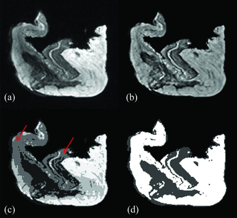
Purpose: Quantification of breast density based on three-dimensional breast MRI may provide useful information for the early detection of breast cancer. However, the field inhomogeneity can severely challenge the computerized image segmentation process. In this work, the effect of the bias field in breast density quantification has been investigated with a postmortem study.
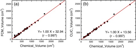
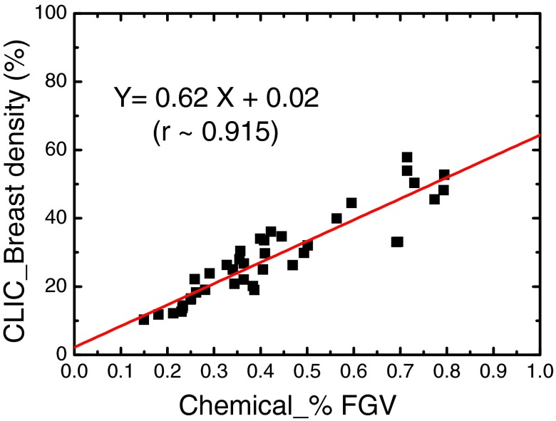
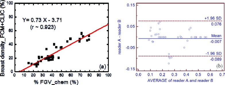
Methods: T1-weighted images of 20 pairs of postmortem breasts were acquired on a 1.5 T breast MRI scanner. Two computer-assisted algorithms were used to quantify the volumetric breast density. First, standard fuzzy c-means (FCM) clustering was used on raw images with the bias field present. Then, the coherent local intensity clustering (CLIC) method estimated and corrected the bias field during the iterative tissue segmentation process. Finally, FCM clustering was performed on the bias-field-corrected images produced by CLIC method. The left-right correlation for breasts in the same pair was studied for both segmentation algorithms to evaluate the precision of the tissue classification. Finally, the breast densities measured with the three methods were compared to the gold standard tissue compositions obtained from chemical analysis. The linear correlation coefficient, Pearson's r, was used to evaluate the two image segmentation algorithms and the effect of bias field.
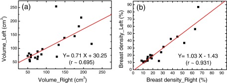
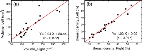
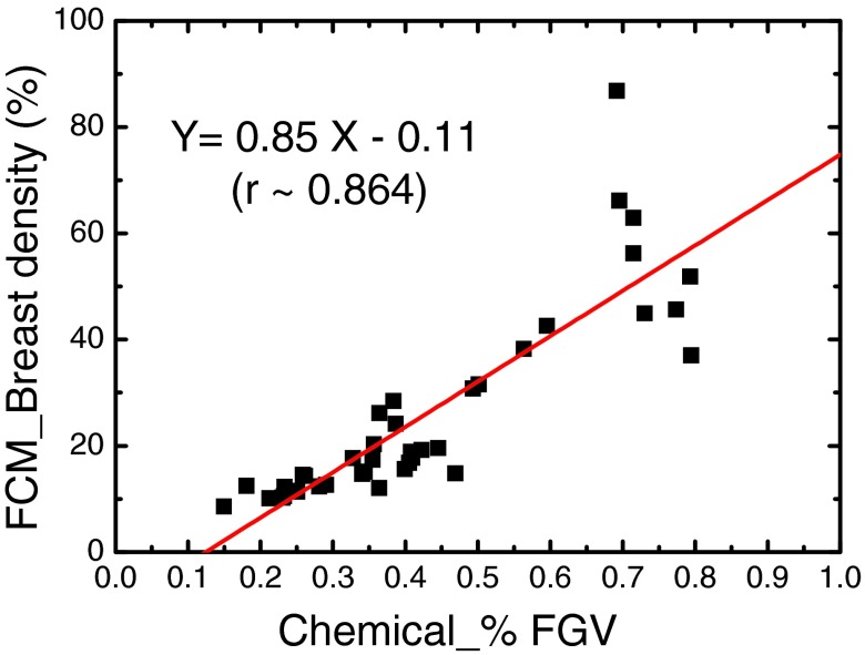
Results: The CLIC method successfully corrected the intensity inhomogeneity induced by the bias field. In left-right comparisons, the CLIC method significantly improved the slope and the correlation coefficient of the linear fitting for the glandular volume estimation. The left-right breast density correlation was also increased from 0.93 to 0.98. When compared with the percent fibroglandular volume (%FGV) from chemical analysis, results after bias field correction from both the CLIC the FCM algorithms showed improved linear correlation. As a result, the Pearson's r increased from 0.86 to 0.92 with the bias field correction.
Conclusions: The investigated CLIC method significantly increased the precision and accuracy of breast density quantification using breast MRI images by effectively correcting the bias field. It is expected that a fully automated computerized algorithm for breast density quantification may have great potential in clinical MRI applications.
Figures
Similar articles
-
A new bias field correction method combining N3 and FCM for improved segmentation of breast density on MRI.Med Phys. 2011 Jan;38(1):5-14. doi: 10.1118/1.3519869. Med Phys. 2011. PMID: 21361169 Free PMC article.
-
Automated fibroglandular tissue segmentation and volumetric density estimation in breast MRI using an atlas-aided fuzzy C-means method.Med Phys. 2013 Dec;40(12):122302. doi: 10.1118/1.4829496. Med Phys. 2013. PMID: 24320533 Free PMC article.
-
Estimation of breast percent density in raw and processed full field digital mammography images via adaptive fuzzy c-means clustering and support vector machine segmentation.Med Phys. 2012 Aug;39(8):4903-17. doi: 10.1118/1.4736530. Med Phys. 2012. PMID: 22894417 Free PMC article.
-
Breast density quantification with cone-beam CT: a post-mortem study.Phys Med Biol. 2013 Dec 7;58(23):8573-91. doi: 10.1088/0031-9155/58/23/8573. Phys Med Biol. 2013. PMID: 24254317 Free PMC article.
-
Fuzzy C-Means Clustering: A Review of Applications in Breast Cancer Detection.Entropy (Basel). 2023 Jul 4;25(7):1021. doi: 10.3390/e25071021. Entropy (Basel). 2023. PMID: 37509968 Free PMC article. Review.
Cited by
-
Postmortem validation of breast density using dual-energy mammography.Med Phys. 2014 Aug;41(8):081917. doi: 10.1118/1.4890295. Med Phys. 2014. PMID: 25086548 Free PMC article.
-
Automatic Estimation of Volumetric Breast Density Using Artificial Neural Network-Based Calibration of Full-Field Digital Mammography: Feasibility on Japanese Women With and Without Breast Cancer.J Digit Imaging. 2017 Apr;30(2):215-227. doi: 10.1007/s10278-016-9922-9. J Digit Imaging. 2017. PMID: 27832519 Free PMC article.
-
Breast density evaluation using spectral mammography, radiologist reader assessment, and segmentation techniques: a retrospective study based on left and right breast comparison.Acad Radiol. 2015 Aug;22(8):1052-9. doi: 10.1016/j.acra.2015.03.017. Epub 2015 May 29. Acad Radiol. 2015. PMID: 26031229 Free PMC article.
-
Predicting Infiltrative Hepatocellular Carcinoma Patient Outcome Post-TACE: MR Bias Field Correction Effect on 3D-quantitative Image Analysis.J Clin Transl Hepatol. 2020 Sep 28;8(3):292-298. doi: 10.14218/JCTH.2020.00054. Epub 2020 Aug 18. J Clin Transl Hepatol. 2020. PMID: 33083252 Free PMC article.
References
-
- Santen R. J., Boyd N. F., Chlebowski R. T., Cummings S., Cuzick J., Dowsett M., Easton D., Forbes J. F., Key T., Hankinson S. E., Howell A., and Ingle J., “Critical assessment of new risk factors for breast cancer: Considerations for development of an improved risk prediction model,” Endocrinol. Relat. Cancer 14, 169–187 (2007).10.1677/ERC-06-0045 - DOI - PubMed
Publication types
MeSH terms
Grants and funding
LinkOut - more resources
Full Text Sources
Other Literature Sources
Medical
Research Materials
