

P-wave abnormality predicts recurrence of atrial fibrillation after electrical cardioversion: a prospective study
- PMID: 24320565
- PMCID: PMC6932521
- DOI: 10.1111/anec.12087
P-wave abnormality predicts recurrence of atrial fibrillation after electrical cardioversion: a prospective study
Abstract
Background: Maintenance of atrial fibrillation (AF) is related to atrial electrical inhomogeneity and resultant chaotic reentry. Our aim was to test the hypothesis that abnormalities of P morphology on the surface electrocardiogram (ECG) predict recurrent AF following electrical cardioversion (ECV).
Methods: A 12-lead ECG was recorded after ECV for persistent AF in 77 patients (51 men, 65 ± 10 years) and repeated 1 month later. P-wave duration was obtained in each lead using blinded on-screen measurement. Maximum P-wave duration (P-max) was defined as the longest measurable P-wave duration in any lead. P-wave dispersion (PWd) was calculated as the maximum-minimum P-wave duration.
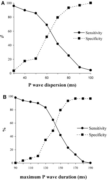
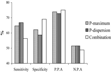
Results: One month after ECV, 29 (38%) patients maintained sinus rhythm. Compared with the sinus rhythm group, those with recurrent AF had significantly greater PWd (66 ± 19 vs 57 ± 16 ms, P = 0.024) and included more patients with P-max ≥142 ms (65% vs 38%, P = 0.023). Using a cutoff of ≥62 ms for PWd and ≥142 ms for P-max, both indices had similar predictive value (sensitivity 66.7 and 64.6%, specificity 58.6 and 62.1%, respectively). In multiple regression analysis, including established clinical predictors, P-max ≥142 ms was the only independent predictor of AF recurrence (P = 0.025).
Conclusion: A prolonged P-wave duration measured by 12-lead ECG predicts recurrent AF within 1 month after ECV.
Keywords: P wave; atrial fibrillation; electrical cardioversion; electrical dispersion; recurrence of atrial fibrillation; surface ECG.
©2013 Wiley Periodicals, Inc.
Figures
Comment in
-
P-wave duration or P-wave morphology? Interatrial block: seeking for the Holy Grail to predict AF recurrence.Ann Noninvasive Electrocardiol. 2014 Jul;19(4):406-8. doi: 10.1111/anec.12156. Epub 2014 May 14. Ann Noninvasive Electrocardiol. 2014. PMID: 24829074 Free PMC article. No abstract available.
-
Response to baranchuk et Al.Ann Noninvasive Electrocardiol. 2014 Jul;19(4):409. doi: 10.1111/anec.12176. Ann Noninvasive Electrocardiol. 2014. PMID: 25040482 Free PMC article. No abstract available.
References
-
- Moe GK. On the multiple wavelet hypothesis of atrial fibrillation. Arch Int Pharmacodyn Ther 1962;140:183–188.
-
- Allessie MA, Lammers WJEP, Bonke FIM, et al. Experimental evaluation of Moe's multiple wavelet hypothesis of atrial fibrillation In Zipes DP. (ed.): Cardiac Electrophysiology and Arrhythmias. New York, Grune & Stratton, 1985, pp. 265–2756.
-
- Haft JI, Lau SH, Stein E, et al. Atrial fibrillation produced by atrial stimulation. Circulation 1968;37:70–74. - PubMed
-
- Spach MS, Miller WT, Geselowitz DB, et al. The discontinuous nature of propagation in normal canine cardiac muscle: Evidence for recurrent discontinuities of intracellular resistance that affect the membrane currents. Circ Res 1981;48:39–54. - PubMed
-
- Gallagher MM, Obel OA, Camm AJ. Tachycardia induced atrial myopathy: An important mechanism in the pathophysiology of atrial fibrillation? J Cardiovasc Electrophysiol 1997;8:1065–1074. - PubMed
Publication types
MeSH terms
Grants and funding
LinkOut - more resources
Full Text Sources
Other Literature Sources
Medical
