. 2013 Dec 15;38(24):5228-5231.
doi: 10.1364/ol.38.005228.

Absolute photoacoustic thermometry in deep tissue

Affiliations

Absolute photoacoustic thermometry in deep tissue

Junjie Yao et al. Opt Lett. .

Abstract

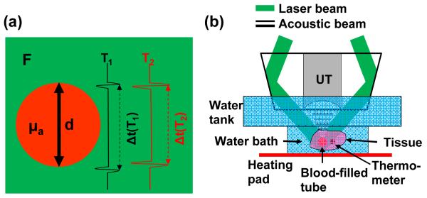
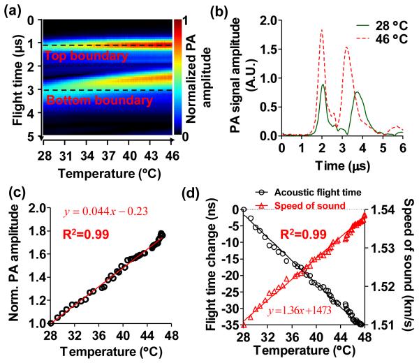
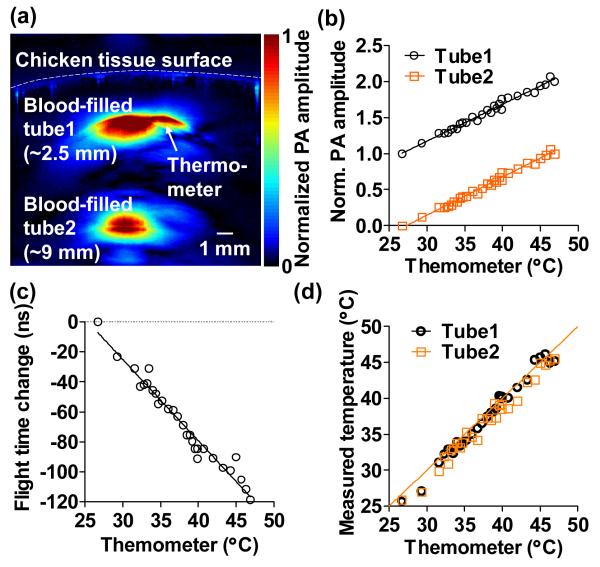
Photoacoustic thermography is a promising tool for temperature measurement in deep tissue. Here we propose an absolute temperature measurement method based on the dual temperature dependences of the Grüneisen parameter and the speed of sound in tissue. By taking ratiometric measurements at two adjacent temperatures, we can eliminate the factors that are temperature irrelevant but difficult to correct for in deep tissue. To validate our method, absolute temperatures of blood-filled tubes embedded ~9 mm deep in chicken tissue were measured in a biologically relevant range from 28°C to 46°C. The temperature measurement accuracy was ~0.6°C. The results suggest that our method can be potentially used for absolute temperature monitoring in deep tissue during thermotherapy.


Figures

Fig. 1
Fig. 1
Absolute temperature measurement by deep photoacoustic macroscopy (PAMac). (a) Illustration of the phantom parameters used for the temperature measurement. F, local laser fluence; μa, absorption coefficient of the blood-filled tube; d, diameter of the blood-filled tube. When the temperature increases from T1 to T2, both the PA signal amplitude and the speed of sound increase. (b) Schematic of the temperature measurement on PAMac. The blood-filled tube and thermometer were embedded in fresh chicken breast tissue suspended in a water bath, whose temperature was regulated by an electrical heating pad and homogenized by continuous stirring. UT, ultrasonic transducer.
Fig. 2
Fig. 2
Systematic calibrations for absolute temperature measurement. (a) Assembly of the time-resolved A-line signals of a 3-mm-diameter blood-filled tube in a clear medium with changing temperature. The top and bottom boundaries of the tube are marked by the dashed lines. As the temperature increased, the PA signal amplitude increased and the acoustic flight time between the two boundaries decreased. The A-lines were aligned at the top boundary for better visualization of the change in acoustic flight time. (b) Representative A-line signals at 28 °C and 46 °C, showing the changes in PA signal amplitude and the acoustic flight time. (c) Normalized PA signal amplitude as a function of temperature between 28 °C to 46 °C, showing the temperature dependence of Grüneisen parameter. Solid line: linear fitting. (d) Change in acoustic flight time (left axis) and speed of sound (right axis) as a function of temperature. Solid lines: first-order rational fitting for flight time, linear fitting for speed of sound.
Fig. 3
Fig. 3
Absolute temperature measurement in deep tissue. (a) Representative cross-sectional image acquired at 800 nm, showing two blood-filled tubes embedded in chicken breast tissue at depths of ~2.5 mm and ~9 mm, respectively. A thermometer was also embedded at the same depth as the top tube. (b). Relative changes in the PA amplitudes from the two tubes followed the same slopes as a function of thermometer readings, which indicated the homogeneous temperature distribution in the tissue. The signals were shifted for clarity. Solid lines: linear fitting. (c) Change in the acoustic flight time between the two tubes as a function of temperature. Solid line: first-order rational fitting. (d) Absolute temperatures measured at the top and bottom tubes as a function of the thermometer readings. Solid line: y = x.

References

    1. Falk MH, Issels RD. Int J Hyperther. 2001;17:1–18. - PubMed
    1. Goldberg SN, Gazelle GS, Mueller PR. Am J Roentgenol. 2000;174:323–331. - PubMed
    1. Dewhirst MW, Viglianti BL, Lora-Michiels M, Hanson M, Hoopes PJ. Int J Hyperther. 2003;19:267–294. - PubMed
    1. Quesson B, de Zwart JA, Moonen CTW. J Magn Reson Imaging. 2000;12:525–533. - PubMed
    1. MaassMoreno R, Damianou CA. J Acoust Soc Am. 1996;100:2514–2521. - PubMed

Publication types